Mesoderm: The Middle Germ Layer of Embryonic Development
In the earliest stages of animal life, the embryo organizes itself into three primary germ layers. The mesoderm is the middle layer, situated between the outer ectoderm and the inner endoderm. This critical layer emerges during a complex process called gastrulation, serving as the biological foundation for many of the body's most vital structural and functional systems.
The development of the mesoderm is driven by intercellular signaling and the influence of an organizing center. This process is regulated by beta-catenin, a co-factor that activates the transcription factor tcf-3 to initiate the synthesis of genes essential for differentiation. Beyond forming its own tissues, the mesoderm is capable of inducing the growth of other structures, such as the neural plate, which eventually becomes the nervous system.
Key Facts
- Formation: Appears during the third week of human embryonic development via gastrulation.
- Primary Components: Divided into axial, paraxial, intermediate, and lateral plate mesoderm.
- Key Derivatives: Forms the heart, blood vessels, muscles, bones, kidneys, and the adrenal cortex.
- Myogenesis: The specific process by which mesenchyme (embryonic connective tissue) forms muscles.
- Structural Role: Creates the notochord, which establishes the anterior-posterior body axis.
The Process of Gastrulation
Gastrulation begins in the third week of human development with the appearance of the primitive streak on the epiblast surface. Through a process called invagination, cells from the epiblast slip beneath the primitive streak. Some of these cells displace the hypoblast to create the endoderm, while others settle between the endoderm and epiblast to form the mesoderm. The remaining cells constitute the ectoderm.
Following this, the chordamesoderm forms the notochord, a central rod that induces the formation of the neural tube and defines the body's primary axis. This sequence of events occurs rapidly: the primitive streak and embryonic mesoderm proliferate between days 13 and 15, the notochord process occurs between days 15 and 17, and the first three somites appear between days 17 and 19.
Classification and Derivatives of the Mesoderm
The mesoderm differentiates into four primary regions, each responsible for specific anatomical structures:
Axial Mesoderm
The axial mesoderm is the central core that gives rise to the notochord, which is essential for signaling the development of the surrounding tissues.
Paraxial Mesoderm
This region organizes into segments. In the head, these are called somitomeres (or neuromeres if they contact the neural plate). In the body, they form somites, which further differentiate into three compartments:
- Sclerotome: Forms cartilage and bone.
- Myotome: Forms skeletal muscle.
- Dermatome: Forms the dermis of the back and subcutaneous tissue.
Intermediate Mesoderm
Connecting the paraxial and lateral plate regions, the intermediate mesoderm develops into the urogenital system, including the kidneys, gonads, associated ducts, and the adrenal cortex.
Lateral Plate Mesoderm
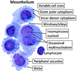
The lateral plate splits into two distinct layers: the parietal (somatic) layer and the visceral (splanchnic) layer. The parietal layer forms the lateral body wall and the mesothelial membranes (serous membranes) lining the pleural, pericardial, and peritoneal cavities. The visceral layer forms the walls of the gut tube. Together, they contribute to the heart, blood vessels, blood cells, and the mesodermal components of the limbs.
Molecular Regulation of Development
The differentiation of somites is controlled by a precise cocktail of proteins and signals from the notochord, neural tube, and epidermis. For example, the proteins SHH (from the notochord and neural tube) and PAX1 are critical for sclerotome (bone/cartilage) formation. Meanwhile, WNT1 and PAX2 drive the creation of the myotome and dermatome.
Bilateral symmetry—ensuring the left and right sides of the body develop identically—is maintained by retinoic acid. This endogenous signal coordinates the segmentation oscillations of the mesoderm, ensuring the vertebral column and other paired structures are aligned.
| Mesoderm Component | Primary Derivatives | Key Structures |
|---|---|---|
| Axial | Notochord | Body axis, neural tube induction |
| Paraxial | Somites | Axial skeleton, skeletal muscle, dermis |
| Intermediate | Urogenital system | Kidneys, gonads, adrenal cortex |
| Lateral Plate | Circulatory & Body Wall | Heart, blood vessels, serous membranes, limb components |
Frequently Asked Questions
What is the difference between the mesoderm and other germ layers?
The mesoderm is the middle layer. The ectoderm is the outermost layer (forming skin and nervous system), and the endoderm is the innermost layer (forming the lining of the gut and respiratory system).
What is myogenesis?
Myogenesis is the biological process of muscle formation, which is specifically a function of the mesenchyme derived from the mesoderm.
How does the notochord influence the embryo?
The notochord establishes the anterior-posterior body axis and sends signals that induce the overlying ectoderm to form the neural tube, the precursor to the central nervous system.
What are somites and why are they important?
Somites are paired blocks of paraxial mesoderm that organize the axial skeleton. They differentiate into the sclerotome (bone), myotome (muscle), and dermatome (skin), ensuring the segmented structure of the vertebrate body.
What role does retinoic acid play in mesoderm development?
Retinoic acid acts as a signal to maintain bilateral synchrony, ensuring that the left and right sides of the embryo develop symmetrically.