Blastulation in Early Animal Embryonic Development
Blastulation is a critical phase in early animal embryonic development that transforms a solid ball of cells into a hollow structure. This process marks the transition from a morula—a solid cluster of cells resulting from the initial cleavage of a fertilized zygote—into a blastula. The blastula serves as the essential architectural foundation for the subsequent stage of development, known as gastrulation, where the primary germ layers of the embryo are established.
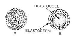
At its core, a blastula is a hollow sphere composed of cells called blastomeres. These cells surround a central, fluid-filled cavity known as the blastocoel. In mammals, this structure is specifically referred to as a blastocyst, which features a more complex organization including a differentiated inner cell mass and an outer layer.
Key Facts
- Timing: In mammals, blastulation typically occurs around day 4 of development.
- Structure: The blastula consists of a blastoderm (cell layer) surrounding the blastocoel (fluid cavity).
- Mammalian Specifics: The blastocyst contains an embryoblast (inner cell mass) for the fetus and a trophoblast for extra-embryonic tissues.
- Control Shift: The Mid-Blastula Transition (MBT) shifts developmental control from maternal mRNA to the embryo's own DNA.
- Cell Adhesion: Cadherins (such as E-cadherin in mammals) are vital for cell polarity and structural integrity.
The Process of Blastulation
Development begins when a sperm fertilizes an egg to create a zygote. This single cell undergoes rapid divisions called cleavages, forming the morula. Blastulation officially begins when the blastocoel first appears. In Xenopus (African clawed frog), this cavity originates from the first cleavage furrow, which widens and is sealed by tight junctions.
The Mid-Blastula Transition (MBT)
In many organisms, including Drosophila (fruit fly) and Xenopus, the early stages of development are governed by maternal mRNA—genetic instructions produced in the egg before fertilization. The Mid-Blastula Transition is a pivotal turning point characterized by:
- Cell Cycle Changes: The synchronous division cycles end, and G1 and G2 growth phases are added. This allows cells to increase in size, ending the period of reductive division.
- Genetic Shift: Maternal mRNA is degraded by proteins (like SMAUG in Drosophila) or microRNA, and the embryo's own genome begins transcribing new mRNA.
Structural Organization
The blastocoel is not merely an empty space; it contains proteins, amino acids, growth factors, ions, and sugars necessary for cellular differentiation. It also provides the physical space required for blastomeres to migrate during gastrulation.
Regional Differences in Xenopus
In Xenopus embryos, the blastula is divided into three distinct regions that determine future tissue types:
- Animal Cap: The roof of the blastocoel, primarily forming ectodermal derivatives.
- Marginal Zone: The walls of the blastocoel, primarily differentiating into mesodermal tissue.
- Vegetal Mass: The floor of the blastocoel, primarily developing into endodermal tissue.
The Mammalian Blastocyst
Mammalian development involves three specific lineages: the epiblast (which becomes the fetus), the trophoblast (which forms part of the placenta), and the primitive endoderm (which becomes the yolk sac). In mice, blastocoel formation begins at the 32-cell stage. This is driven by sodium-potassium pumps that create an osmotic gradient, drawing water into the embryo via aquaporins.
| Feature | Mammals (Blastocyst) | Amphibians (Xenopus) |
|---|---|---|
| Key Cell Types | Epiblast, Trophoblast, Primitive Endoderm | Animal Cap, Marginal Zone, Vegetal Mass |
| Primary Adhesion Molecule | E-cadherin | EP-cadherin / XB/U cadherin |
| Cavity Formation | Osmotic gradient via Na+/K+ pumps | Widening of the first cleavage furrow |
| Outcome | Fetus and extra-embryonic tissues | Ectoderm, Mesoderm, and Endoderm |
Cellular Adhesion and Polarity
The establishment of cell polarity (the spatial difference in shape and function across a cell) is essential for development. This is achieved through tight junctions and cadherin-mediated interactions. In mammals, a process called compaction occurs around the 8-cell stage, where E-cadherins and catenins transform a loose group of cells into a polarized, cohesive ball.
These junctions create a permeability seal that regulates the blastocoel's environment and allows for paracellular transport, setting the stage for the embryo to transition from an indistinct mass to a structured organism.
Clinical and Research Implications
Assisted Reproductive Technology
Research into preimplantation embryos has led to advancements in in vitro fertilization (IVF). Studies in mice have demonstrated that hormonal induction, superovulation, and artificial insemination can successfully produce embryos for uterine implantation, providing a framework for human reproductive medicine.
Stem Cell Research and Regenerative Medicine
Cells at the blastula stage are often pluripotent, meaning they have the potential to become almost any cell type in the body. By manipulating cell signaling and transcription factors, scientists can direct these cells toward specific lineages. For example, pluripotent Xenopus cells have been successfully induced to form functional retinas when transplanted to the neural plate, offering significant potential for treating injuries and degenerative diseases.
Frequently Asked Questions
What is the difference between a morula and a blastula?
A morula is a solid ball of cells produced by early cleavage, while a blastula is a hollow sphere of cells that has developed a fluid-filled cavity called the blastocoel.
What happens during the Mid-Blastula Transition?
The Mid-Blastula Transition is when the embryo stops relying on maternal mRNA and begins transcribing its own DNA. It also marks the point where cell cycles lengthen to include growth phases (G1 and G2), allowing the embryo to increase in overall size.
What is the role of the trophoblast in mammals?
The trophoblast is the outer layer of the mammalian blastocyst that eventually develops into the extra-embryonic tissues, including part of the placenta.
Why are blastula cells important for regenerative medicine?
Because blastula cells are pluripotent, they can be directed to become specific organ-specific cells. This ability to create functional tissues, such as retinas, makes them invaluable for researching ways to repair damaged organs or treat diseases.
How is the blastocoel formed in mammalian embryos?
It is formed through an osmotic gradient created by sodium-potassium pumps in the trophectoderm, which causes water to enter the embryo through channels called aquaporins.