Invagination: The Cellular Mechanics of Tissue Folding in Development
In the complex choreography of embryonic development, invagination serves as a fundamental mechanical process where a surface folds inward to create a cavity, pouch, or tube. This biological phenomenon is essential for shaping the architecture of an organism, playing a critical role in the formation of the primitive gut during gastrulation, the development of the neural tube in vertebrates, and the morphogenesis of various sensory structures and organs.
While invagination occurs across a diverse array of species, the core principle remains the same: the mechanics of one side of an epithelial sheet (a layer of cells) are altered, creating pressure that induces a bend in the tissue.
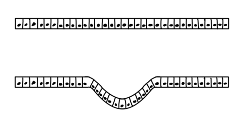
Key Facts
- Definition: The process of a surface folding in on itself to form a internal cavity or tube.
- Primary Driver: Often powered by the actin-myosin cytoskeleton, which generates contractile force.
- Cell Shape Change: Cells typically transition from cube or column shapes to wedge shapes to facilitate bending.
- Critical Roles: Essential for gastrulation (gut formation) and neurulation (spinal cord precursor formation).
- Diversity: While apical constriction is common, some tissues use basal constriction or supracellular cables to fold.
Cellular Mechanisms of Invagination
Regardless of the specific force used, most invagination events result in a stereotypical change in cell geometry. In a typical scenario, the apical side (the surface exposed to the environment) shrinks, while the basal side (the side contacting the basement membrane) expands. This transforms the cells into wedge shapes, forcing the tissue to bend toward the apical surface.
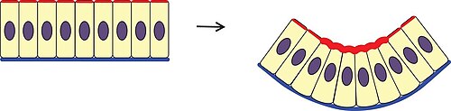
Apical Constriction
Apical constriction is an active process where the apical surface of a cell shrinks. This is powered by the actin-myosin cytoskeleton, a network where the motor protein myosin pulls actin filaments together. This activity is regulated by the phosphorylation of the myosin regulatory light chain, managed by kinases like Rho-associated coiled-coil kinase (ROCK) and various phosphatases.
Basal Relaxation and Height Changes
To maintain a constant volume while the top shrinks, cells must either elongate or expand their base. Basal relaxation involves the active disassembly of the actin-myosin network at the basal surface, allowing it to expand. This has been observed during the formation of the otic placode in chickens and the ventral furrow in Drosophila.
Additionally, changes in cell height can drive folding. In some cases, cells elongate, thickening the epithelium. In others, cells shorten; for example, in ascidian gastrulation, cells become rounder after apical constriction to complete the invagination. In the Drosophila tracheal placode, the rounding of cells during mitosis (cell division) contributes to the process.
Supracellular Cables
Not all folding relies on individual cell constriction. Supracellular actomyosin cables are networks of actin and myosin that span multiple cells, connected by cell junctions. These cables can contract around a site to create compressive forces, as seen in the salivary gland invagination of Drosophila, or pull tissue together to facilitate tube formation in the chick embryo's neural tube.
Notable Biological Examples
Drosophila Ventral Furrow
In Drosophila melanogaster, the prospective mesoderm along the ventral midline folds inward to create the ventral furrow. This is one of the first major movements of gastrulation. The process is genetically controlled by transcription factors twist and snail, with the Fog signaling pathway regulating the apical changes in the cells.
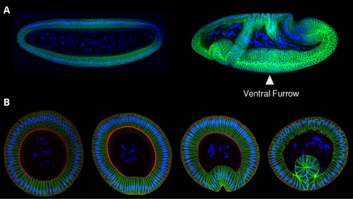
Neural Tube Formation
Primary neurulation is the process by which the neural plate invaginates to form the neural tube, the precursor to the spinal cord. This involves a medial hinge point where tissue bending begins. In Xenopus frogs, this is driven by apical constriction. However, in mice and chickens, it occurs via basal wedging, where the nucleus moves to the basal side, creating a bulge. Supracellular cables and external pressure from adjacent tissues also assist in closing the tube.
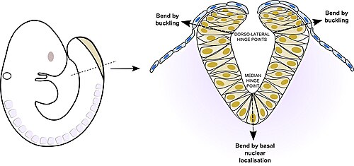
Sea Urchin and Amphioxus Gastrulation
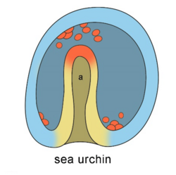
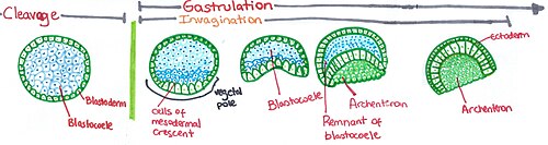
In sea urchins, the vegetal plate invaginates to form the archenteron (the future gut). This occurs in stages: initial folding, elongation of the tube, and finally, contact with the opposite side of the cell cavity. Similar processes of invagination are observed during the gastrulation of amphioxus.
Summary of Invagination Mechanisms
| Mechanism | Primary Action | Key Driver/Protein | Example |
|---|---|---|---|
| Apical Constriction | Shrinkage of apical surface | Actin-myosin network | Xenopus neural tube |
| Basal Wedging | Expansion of basal surface | Nuclear positioning | Mouse/Chick neural tube |
| Supracellular Cables | Coordinated tissue pulling | Intercellular actomyosin cables | Drosophila salivary glands |
| Cell Rounding | Reduction in cell height | Mitosis / Apoptosis | Drosophila tracheal placode |
Other Forms of Invagination
Beyond embryonic morphogenesis, invagination occurs at the cellular level during endocytosis and exocytosis, where the cell membrane folds inward to form vesicles. In medical contexts, the invagination of one part of the intestine into another is known as intussusception.
Frequently Asked Questions
What is the difference between apical constriction and basal wedging?
Apical constriction involves the shrinking of the cell's top surface to create a wedge shape, whereas basal wedging involves the expansion of the bottom surface, often caused by the movement of the nucleus to the basal side.
What role does myosin play in tissue folding?
Myosin acts as a motor protein that pulls actin filaments together. This contraction creates the physical force necessary to shrink the apical surface of cells, leading to the bending of the epithelial sheet.
How does the ventral furrow form in Drosophila?
It is driven by the transcription factors twist and snail, which trigger the Fog signaling pathway. This leads to apical constriction of the cells along the ventral midline, folding them inward.
Is invagination only found in embryos?
No. While critical for embryos, invagination also occurs during basic cellular functions like endocytosis (forming vesicles) and can occur pathologically in the intestines (intussusception).
What are supracellular cables?
These are actin-myosin structures that align across multiple adjacent cells. Instead of individual cells shrinking, these cables contract across the tissue to pull it into a fold or tube.