Ectoderm: The Outermost Germ Layer and Its Role in Development
In the earliest stages of embryonic development, a complex biological process organizes a cluster of cells into three primary germ layers. The ectoderm is the outermost of these layers, positioned superficially to the mesoderm (the middle layer) and the endoderm (the innermost layer). Derived from the Greek words ektos (outside) and derma (skin), the ectoderm serves as the foundational blueprint for the body's external interfaces and the complex systems that allow an organism to interact with its environment.
Generally, the ectoderm differentiates into epithelial and neural tissues. This includes the skin, the linings of the nostrils, mouth, and anus, as well as hair, nails, sweat glands, and tooth enamel. It is also responsible for the creation of the brain, spinal cord, and nerves.
Key Facts
- Position: The outermost of the three primary germ layers.
- Primary Derivatives: Forms the nervous system (brain, spinal cord) and the epidermis (skin, hair, nails).
- Subdivisions: Divided into the surface ectoderm and the neuroectoderm (neural plate and neural crest).
- Key Processes: Develops through gastrulation and neurulation.
- Clinical Relevance: Mutations affecting this layer can lead to ectodermal dysplasia.
The History of Germ Layer Discovery
The discovery of the three germ layers is credited to Heinz Christian Pander, a Baltic German-Russian biologist. Using chicken eggs for his research, Pander identified the ectoderm, mesoderm, and endoderm, earning him a reputation as the founder of embryology.
This work was expanded by Karl Ernst von Baer, a Prussian-Estonian biologist. Baer extended Pander's principles to all vertebrates and discovered the blastula (an early hollow ball of cells). In 1828, Baer published these findings in his seminal textbook, On the Development of Animals.
The Process of Differentiation
Initial Appearance and Early Development
In fish and amphibians, the ectoderm first appears during the later stages of gastrulation. The embryo begins as a polar blastula, divided into an animal hemisphere and a vegetal hemisphere; the animal hemisphere eventually becomes the ectoderm.
The positioning of the ectoderm is governed by selective affinity. The inner surface of the ectoderm has a strong positive affinity for the mesoderm and a weak negative affinity for the endoderm. This attraction is regulated by cadherin molecules on the cell surfaces. For instance, N-cadherin is essential for separating precursor neural cells from precursor epithelial cells.
Gastrulation and Layer Formation
During gastrulation, bottle cells invaginate on the dorsal surface of the blastula to form the blastopore, creating a fluid-filled cavity called the blastocoel. Through processes known as radial extension and convergent extension (where cells intercalate mediolaterally), the prospective mesoderm is positioned between the ectoderm and endoderm.
Simultaneously, the ectoderm cells undergo epiboly, dividing to form a single uniform layer that completely engulfs the vegetal pole (the future endoderm), resulting in a three-layered embryo.
Neurulation and Organogenesis
The Formation of the Nervous System
Once the layers are established, neurulation begins. This is the process by which the ectoderm differentiates into the epidermis, the neural crest, and the neural tube. This occurs in two phases:
- Primary Neurulation: The notochord signals the superficial ectoderm to form the neural plate. Medial hinge cells (MHPs) and dorsolateral hinge cells (DLHPs) create a wedge-like folding effect, causing the plate to converge.
- Secondary Neurulation: This further positions the neural crest cells between the deep neural tube and the superficial epidermal layer.
The resulting structures have distinct fates: the neural tube forms the central nervous system, the neural crest forms the peripheral and enteric nervous systems, facial cartilage, and melanocytes, and the epidermis forms the skin, hair, nails, and eyes.
Organogenesis and Molecular Signaling
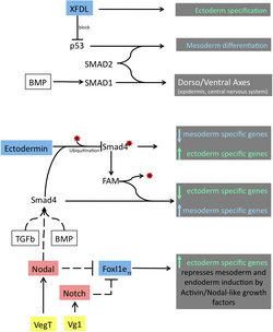
Ectodermal organs originate from the interaction between the epithelium and the mesenchyme. This process is mediated by signaling molecules including FGF, TGFβ, Wnt, and the hedgehog family. In tooth development, FGF-9 increases the rate of epithelial invagination, while FGF-10 stimulates cell proliferation to enlarge the tooth germs.
Mammalian teeth specifically develop from oral ectoderm and neural crest cells, with stem cells for continuous growth forming from the suprabasal layer of the surface ectoderm and the stellate reticulum.
Clinical Significance: Ectodermal Dysplasia
Ectodermal dysplasia refers to a group of over 170 rare genetic conditions where tissues derived from the ectoderm develop abnormally. These conditions typically affect the skin, hair, nails, sweat glands, and teeth.
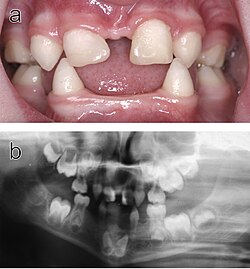
The most common subtype is Hypohidrotic Ectodermal Dysplasia (HED). A primary symptom is hypohidrosis—the inability to sweat sufficiently due to missing or dysfunctional sweat glands. This can lead to life-threatening hyperthermia in warm climates. Other symptoms include conical or absent teeth, thin hair, and facial malformations.
HED is often linked to variants of the X-chromosomal EDA gene. Because it is X-linked, the condition typically affects males more severely than females.
| Ectoderm Division | Primary Derivatives | Key Signaling/Factors | Associated Pathology |
|---|---|---|---|
| Surface Ectoderm | Epidermis, hair, nails, tooth enamel, oral epithelium | FGF-9, FGF-10 | Ectodermal Dysplasia (HED) |
| Neural Tube | Central Nervous System (Brain, Spinal Cord) | Notochord signaling | Neural Tube Defects |
| Neural Crest | Peripheral nervous system, facial cartilage, melanocytes | N-cadherin | Craniofacial abnormalities |
Frequently Asked Questions
What is the difference between surface ectoderm and neuroectoderm?
The surface ectoderm primarily gives rise to epithelial tissues such as the skin, hair, and nails. The neuroectoderm, which consists of the neural plate and neural crest, gives rise to the majority of the nervous system, including the brain and spinal cord.
How does the ectoderm form during gastrulation?
The ectoderm forms from the animal hemisphere of the blastula. Through a process called epiboly, these cells divide and spread to form a single outer layer that encapsulates the developing mesoderm and endoderm.
What is the role of cadherins in ectoderm development?
Cadherins are molecules on the cell surface that determine the "selective affinity" between germ layers. For example, N-cadherin is crucial for ensuring that precursor neural cells remain separate from precursor epithelial cells.
What happens in Hypohidrotic Ectodermal Dysplasia (HED)?
HED is a genetic condition, often involving the EDA gene, that results in the abnormal development of ectodermal structures. This leads to symptoms such as a lack of sweat glands (hypohidrosis), sparse hair, and missing or pointed teeth.
Which signaling molecules are most important for ectodermal organogenesis?
Key regulators include FGF (Fibroblast Growth Factor), TGFβ (Transforming Growth Factor beta), Wnt, and the hedgehog family. Specifically, FGF-9 and FGF-10 are critical for the initiation and growth of tooth germs.