Understanding Virology: The Science of Biological Viruses
Virology is the specialized scientific study of biological viruses. As a vital subfield of microbiology, it explores the entire lifecycle and impact of these microscopic entities—from their complex structures and evolutionary history to the sophisticated ways they infect and exploit host cells for reproduction.
Beyond basic biology, virology is an essential applied science. It plays a critical role in medical microbiology, veterinary medicine (known as veterinary virology), and plant pathology. By studying viral pathogenesis—the process by which viruses cause disease—scientists can develop better treatments, vaccines, and diagnostic tools to protect human, animal, and plant health.

The Origins of Virology
The field of virology emerged as a distinct discipline from bacteriology in 1898. This breakthrough occurred when Martinus Beijerinck identified the causative agent of tobacco mosaic disease (TMV). Beijerinck realized the pathogen was neither a bacterium nor a fungus, but something entirely different. He famously described this mysterious agent as contagium vivum fluidum, or "contagious living fluid," and used the term "virus" to define it.

Decades later, the scientific understanding of these entities deepened significantly. In 1955, Rosalind Franklin proposed the full structure of the tobacco mosaic virus, providing a foundational piece of the puzzle in structural virology.
How Scientists Detect and Study Viruses
Because viruses are too small to be seen with traditional light microscopes, researchers rely on advanced technologies to identify, isolate, and analyze them.
Visualizing the Invisible: Electron Microscopy
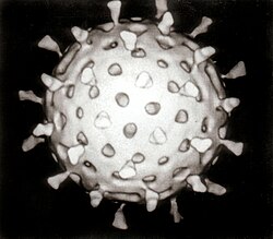
Electron microscopy is a cornerstone of virology, allowing scientists to observe the physical structure of virus particles. This technology has enabled the visualization of various viral types, including rotaviruses, adenoviruses, noroviruses, and astroviruses.


Advanced techniques like cryoelectron microscopy provide even higher resolution, offering detailed views of viral components at a near-atomic level.
Laboratory Detection and Diagnostics
To identify infections, virologists use several sophisticated methods:
- Serology: Studying how the immune system responds to viruses through antibodies.
- Nucleic Acid Detection: Using methods like Polymerase Chain Reaction (PCR) to find viral DNA or RNA.
- Infectivity Assays: Testing how well a virus can actually infect host cells.
- Immunofluorescence: Using fluorescent markers to see viruses within infected cells.

When viruses infect cells, they often cause visible changes known as the cytopathic effect. For example, herpes simplex virus can cause infected cells to become round and balloon-like, or create "plaques" (clear areas) in stained cell cultures.


Purification and Analysis
To study a virus in isolation, scientists must separate it from other biological material. Ultracentrifugation is a primary tool for this. While standard centrifuges are too weak, ultracentrifuges spinning at speeds up to 100,000 rpm can concentrate viruses through differential centrifugation—a process where contaminants are removed at low speeds, and the light, small virus particles are concentrated at high speeds.
![Caesium chloride (CsCl) solution and two morphological types of rotavirus. Following centrifugation at 100,000 g a density gradient forms in the CsCl solution and the virus particles separate according to their densities. The tube is 10 cm tall. The viruses are the two "milky" zones close together.[61]](/images/ad/d7/add7f5a6375a383d7183ea95d4660f68b0c237dedeffac61e93c7562f0e15d4d.jpg)
Once purified, scientists use electrophoresis to separate viral proteins and sequencing to map their entire genetic code. This allows for phylogenetic analysis—studying the evolutionary relationships between different viruses.

Classifying the Virosphere
Because of their immense diversity, viruses are organized into complex hierarchical systems. The International Committee on Taxonomy of Viruses (ICTV) manages a classification system that ranges from the broad Realm down to the specific Species.
The Baltimore Classification
One of the most important ways to categorize viruses is the Baltimore Classification. This system groups viruses based on how they produce messenger RNA (mRNA), which is the template used to make proteins.
| Group | Type of Genetic Material | Examples |
|---|---|---|
| I | Double-stranded DNA (dsDNA) | Adenoviruses, Herpesviruses, Poxviruses |
| II | Single-stranded DNA (ssDNA) | Parvoviruses |
| III | Double-stranded RNA (dsRNA) | Reoviruses |
| IV | Positive-sense single-stranded RNA ((+)ssRNA) | Coronaviruses, Picornaviruses |
| V | Negative-sense single-stranded RNA ((-)ssRNA) | Orthomyxoviruses, Rhabdoviruses |
| VI | ssRNA with DNA intermediate (ssRNA-RT) | Retroviruses |
| VII | dsDNA with RNA intermediate (dsDNA-RT) | Hepadnaviruses |

Key Facts
- Virology is the study of viruses, including their structure, evolution, and how they cause disease.
- Martinus Beijerinck is credited with founding virology in 1898.
- Electron microscopy is required to visualize the physical structure of viruses.
- Ultracentrifugation is used to purify and concentrate virus particles.
- The Baltimore Classification organizes viruses by their method of mRNA synthesis.
Frequently Asked Questions
What is the difference between virology and bacteriology?
Bacteriology is the study of bacteria, which are single-celled living organisms. Virology is the study of viruses, which are distinct from bacteria and require a host cell to reproduce.
How do scientists see viruses if they are so small?
Scientists use electron microscopes, which use beams of electrons rather than light to create highly detailed images of microscopic structures like viruses.
What does "pathogenesis" mean in virology?
Viral pathogenesis refers to the specific biological mechanisms and processes by which a virus infects a host and causes disease.
Why are there different groups in the Baltimore Classification?
Viruses use different genetic strategies to replicate. The Baltimore Classification groups them based on whether they use DNA or RNA, and whether that material is single-stranded or double-stranded, to explain how they produce mRNA.
What is a cytopathic effect?
A cytopathic effect refers to the visible structural changes or damage caused to host cells as a result of a viral infection.