The Microscopic World: An Exploration of Bacteria
Bacteria are among the most ancient and diverse forms of life on Earth. Belonging to the domain Bacteria, these single-celled prokaryotes—organisms that lack a defined nucleus—have existed for an estimated 3.5 billion years, dating back to the Paleoarchean era. From the deepest ocean trenches to the vacuum of space, they have adapted to nearly every environment imaginable.

Classification and Evolutionary Diversity
The biological classification of bacteria is incredibly complex. While they all belong to the same domain, they are divided into numerous kingdoms and phyla. Modern genomic analyses have identified several major supergroups, such as the Bacillati, Fusobacteriati, and Pseudomonadoti. Scientists also recognize many "candidate phyla," which represent groups of bacteria that are yet to be fully characterized.
![Phylogenetic tree of Bacteria, Archaea and Eukarya, with the last universal common ancestor (LUCA) at the root[23]](/images/f1/ef/f1efbbbeb2df62dcb3e208a44eb5501910ac87cda8da3795faae522ef25ea9f1.webp)
The sheer diversity of the bacterial domain is reflected in its vast phylogenetic tree. Recent studies suggest that bacteria represent a massive spectrum of life, including groups like the CPR ultramicrobacterias and the Gracilicutes. This evolutionary complexity highlights how bacteria have branched out to occupy every available ecological niche.
![Phylogenetic tree showing the diversity of bacteria, compared to other organisms. Here bacteria are represented by three main supergroups: the CPR ultramicrobacterias, Bacillati and Gracilicutes according to 2019 genomic analyses[185]](/images/65/60/65605ee56cce7242ff9a866f577dd1a1ae9f2d0a4506bf32cc1c74e1b23991c6.png)
Morphology and Physical Characteristics
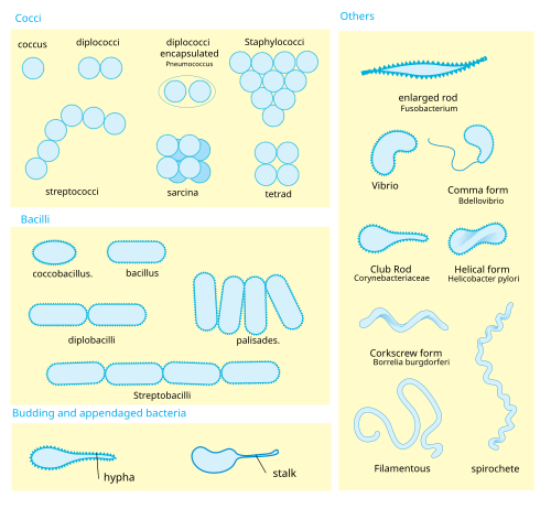
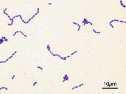
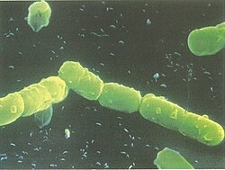
Bacteria are not uniform in appearance; they exhibit a wide variety of shapes and arrangements. Some are spherical, some are rod-shaped, and others are spiral. This diversity in morphology (the study of the form and structure of organisms) is a key factor in how they survive in different environments.
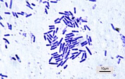
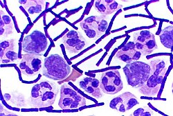
For example, the genus Bacillus is well-known for its rod-shaped structure. These shapes are not merely aesthetic; they are functional adaptations that influence how a bacterium moves, feeds, and interacts with its surroundings.
Size also varies significantly across the bacterial world. While most are microscopic, the range of sizes is vast when compared to other biomolecules and larger organisms. This scale allows them to navigate environments ranging from the tiny spaces between human cells to the vastness of soil ecosystems.
![The range of sizes shown by prokaryotes (Bacteria), relative to those of other organisms and biomolecules[62]](/images/48/7a/487ae7461172cde46eb0ee5af7184eebb85e59b6fc178b5d10b25fce8ca00846.webp)
Cellular Structure and Staining
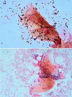
One of the most important ways scientists identify bacteria is through Gram staining, a method used to classify them based on the structure of their cell walls. This process distinguishes between two major types: Gram-positive and Gram-negative bacteria.
Gram-positive bacteria, such as those in the genus Bacillus, possess a thick peptidoglycan layer and typically only one cell membrane. This structure provides a different level of protection and interaction compared to their counterparts.
In contrast, Gram-negative bacteria, such as those in the genus Pseudomonas, have a more complex exterior, often including an additional outer membrane. This structural difference is a critical factor in how these organisms respond to environmental stressors and antibiotics.
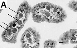
Beyond the cell wall, some bacteria contain specialized internal structures. For instance, certain species utilize carboxysomes—protein-based organelles that help compartment metabolic processes, such as carbon fixation, within the cell.
Movement and Locomotion
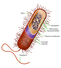
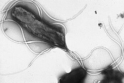
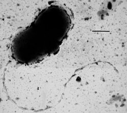
To navigate their environments, many bacteria have developed sophisticated movement mechanisms. The most common method involves flagella, which are whip-like appendages that act like tiny propellers. The arrangement of these flagella can vary, including monotrichous (a single flagellum), lophotrichous (a tuft), or peritrichous (distributed all over the cell surface).

Different species utilize these appendages in unique ways. For example, Helicobacter pylori uses multiple flagella to move through viscous environments, while other species like Desulfovibrio vulgaris may possess a single flagellum at one end of the cell.
Metabolism and Nutrition
Bacteria are incredibly resourceful, utilizing various methods to obtain energy and carbon. They are categorized into three primary nutritional types based on their metabolic needs:
- Phototrophs: These organisms use sunlight as their primary energy source. They can be photoautotrophs, which fix carbon dioxide, or photoheterotrophs, which use organic compounds.
- Lithotrophs: These bacteria derive energy from inorganic compounds. They can be lithoautotrophs (fixing carbon) or lithoheterotrophs (using organic compounds).
- Organotrophs: These organisms rely on organic compounds for both energy and carbon.
| Nutritional Type | Source of Energy | Source of Carbon | Examples |
|---|---|---|---|
| Phototrophs | Sunlight | Organic compounds or Carbon fixation | Cyanobacteria, Green sulfur bacteria |
| Lithotrophs | Inorganic compounds | Organic compounds or Carbon fixation | Thermodesulfobacteriota |
| Organotrophs | Organic compounds | Organic compounds or Carbon fixation | Bacillus, Clostridium |
Survival in Extreme Environments
Bacteria are famous for being extremophiles—organisms that thrive in conditions that would be lethal to most other life forms. Their ability to inhabit extreme niches is a testament to their biological resilience.
Some species thrive in extreme cold, such as those found in Antarctica, while others are thermophiles, living in geysers or submarine hydrothermal vents at temperatures between 70 and 121 °C. Others can survive intense radiation, high salinity, extreme pH levels (from highly acidic to highly alkaline), and even the high pressure of the Mariana Trench. Remarkably, some bacteria have even survived for years in space on NASA satellites.
Reproduction and Growth
Most bacteria reproduce through binary fission, a form of asexual reproduction where a single cell divides into two identical daughter cells. This process is highly efficient and allows for rapid population growth under favorable conditions.
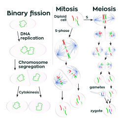
Bacterial growth typically follows a specific pattern known as a growth curve, which includes distinct phases such as the lag phase, the exponential growth phase, and the stationary phase. Understanding these curves is essential for studying how bacterial populations expand and stabilize in different environments.

In some biological interactions, bacteria are even targeted by viruses known as phages. A T4 phage, for example, can infect E. coli by attaching to the cell and injecting its DNA, effectively hijacking the bacterium's machinery.
![Helium ion microscopy image showing T4 phage infecting E. coli. Some of the attached phage have contracted tails indicating that they have injected their DNA into the host. The bacterial cells are ~ 0.5 μm wide[142]](/images/ce/0d/ce0d40726e6dde32089e7a5020db4517b6ace054eb70e46fa3ad580f24da571a.jpg)
Interactions with Other Organisms
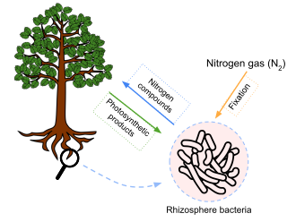
Bacteria play diverse roles in the ecosystems they inhabit. They can exist in mutualistic relationships, where both organisms benefit, such as the nitrogen-fixing bacteria that live in the rhizosphere of plants. However, they can also be pathogens, causing disease in humans and other organisms.
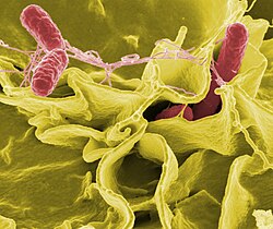
Bacterial infections can manifest in many ways, ranging from common respiratory issues to severe systemic diseases. Pathogens like Bacillus anthracis or Salmonella typhimurium can invade host cells and cause significant harm.
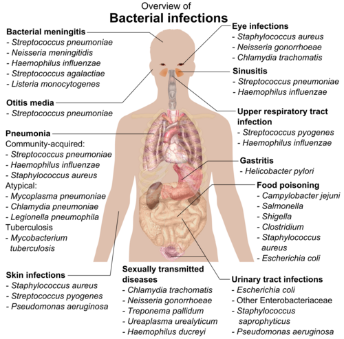
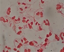
In the human body, the balance of bacteria is crucial. For instance, certain bacteria are part of the normal flora, while others, like Neisseria gonorrhoeae or Salmonella, can lead to infection. Even the displacement of beneficial bacteria, as seen in bacterial vaginosis, can lead to health complications.
Key Facts
- Age: Bacteria have existed for approximately 3.5 billion years.
- Cell Type: They are prokaryotes, meaning they lack a nucleus.
- Reproduction: Most reproduce via binary fission.
- Diversity: They inhabit extreme environments, including high-pressure trenches and outer space.
- Classification: They are categorized by shape, Gram stain reaction, and metabolic type.
Frequently Asked Questions
What are bacteria?
Bacteria are single-celled, prokaryotic organisms that belong to the domain Bacteria. They are found in almost every environment on Earth and can be beneficial, neutral, or harmful to other life forms.
How do bacteria reproduce?
Most bacteria reproduce through a process called binary fission, where a single cell replicates its DNA and divides into two identical cells.
Can bacteria live in extreme environments?
Yes, many bacteria are extremophiles. They can survive in extreme heat, intense cold, high radiation, extreme pH levels, and even the high pressure of the deep ocean.
Are all bacteria harmful to humans?
No. While some bacteria are pathogens that cause diseases, many others are beneficial, living in our bodies as commensals or participating in mutualistic relationships that support health and ecosystems.
What is the difference between Gram-positive and Gram-negative bacteria?
The difference lies in their cell wall structure. Gram-positive bacteria have a thick peptidoglycan layer and a single membrane, while Gram-negative bacteria have a more complex structure, often including an outer membrane.