Skull Anatomy: Structure, Development, and Clinical Significance
The skull is a sophisticated biological structure designed primarily to protect the brain and support the sensory organs of the face. In humans, it serves as a rigid framework that balances extreme durability with the flexibility required for prenatal development and birth. From the complex arrangement of its 22 primary bones to the specialized processes of ossification, the skull is a marvel of evolutionary engineering.
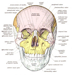
The Structure of the Human Skull
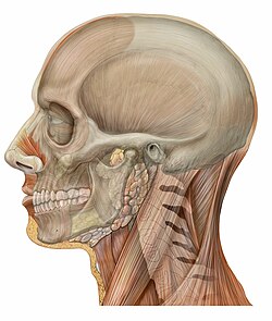
The human skull is generally composed of 22 bones, which are divided into two main functional groups: the neurocranium (the braincase) and the facial skeleton.
The Neurocranium
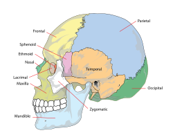
The neurocranium consists of eight bones that enclose and protect the brain. These include:
- The frontal bone
- Two parietal bones
- Two temporal bones
- The occipital bone
- The sphenoid bone
- The ethmoid bone
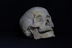
The Facial Skeleton
The facial skeleton comprises 14 bones that form the structure of the face and provide attachment points for muscles. These include:
- Two nasal bones and two lacrimal bones
- Two maxillae (upper jaw) and the mandible (lower jaw)
- Two zygomatic bones (cheekbones)
- Two palatine bones and the vomer
- Two inferior nasal conchae
While 22 is the general consensus, some anatomical counts vary. Some sources include the hyoid bone or the three ossicles of the middle ear—the malleus, incus, and stapes—in the total count.
Key Facts
- Bone Count: The adult human skull typically consists of 22 bones.
- Protection: The skull is one of nature's least deformable structures, requiring approximately 1 ton of force to reduce its diameter by 1 cm.
- Development: Newborns have 44 separate bony elements that fuse over time.
- Ossification: Skull bones form via two processes: intramembranous (dermal) and endochondral ossification.
- Immunity: Tiny channels in the skull allow immune cells from bone marrow to reach brain inflammation sites.
Skull Development and Growth
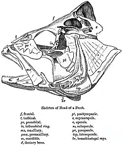
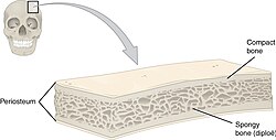
The formation of the skull involves two types of ossification—the process of bone formation. The skull roof and facial bones are dermal bones formed by intramembranous ossification. In contrast, the endocranium (the bones supporting the brain, such as the occipital, sphenoid, and ethmoid) is largely formed by endochondral ossification, where cartilage is replaced by bone.
During the first trimester of pregnancy, the anterior cranial fossa undergoes rapid allometric growth, increasing from 5 to 17 millimeters between the 8th and 14th week. This period is critical, as defects can often develop during this rapid change.
Fontanelles and Sutures
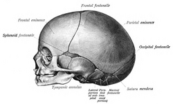
At birth, the skull is not a single solid piece but consists of 44 separate elements. The roof bones are separated by fontanelles—regions of dense, moveable connective tissue. There are six fontanelles: one anterior, one posterior, two sphenoid, and two mastoid. The anterior fontanelle, often called the "soft spot," can remain open for up to 18 months.
As the child grows, these fontanelles are replaced by sutures (fibrous joints). The five primary sutures are the coronal, sagittal, lambdoid, and the two squamous sutures.
Clinical Significance and Medical Advancements
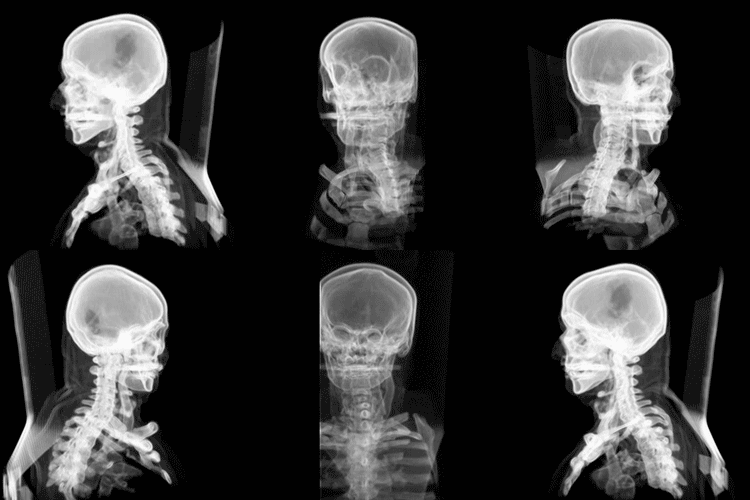
Because the skull is so rigid, it protects the brain effectively but can create dangerous conditions if internal pressure rises. A subdural haematoma (blood clot) can increase intracranial pressure, potentially leading to "coning," where the brain is forced out of the foramen magnum, which can be fatal without urgent surgery.
Modern medicine has introduced groundbreaking treatments for skull injuries and diseases. In 2013, the first 3D-printed polymer implants were used in the U.S. to replace missing sections of the skull. Similarly, a Dutch woman received a complete 3D-printed cranium replacement to treat hyperostosis, a condition where excessive bone thickness compresses the brain.
Comparative Anatomy and Craniometry
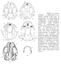
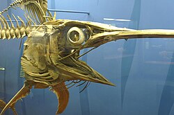
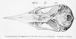
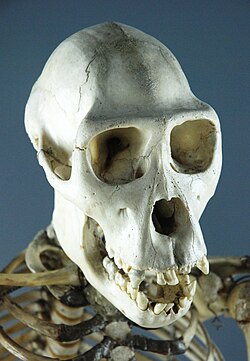
The skull varies significantly across the animal kingdom, from the streamlined skulls of swordfish and cuckoos to the robust structures of chimpanzees and amphibians.
Sexual Dimorphism
In adults, male skulls are generally larger and more robust, while female skulls are lighter and smaller, with a cranial capacity roughly 10% less than males. However, some research suggests women's skulls may be slightly thicker in certain areas, and men may maintain bone density better with age.
Craniometry
Scientists use the cephalic index (the ratio of head width to length) to categorize head shapes:
- Dolichocephalic: Long-headed
- Mesaticephalic: Medium-headed
- Brachycephalic: Short-headed
A vertical cephalic index is also used to classify heads as chamaecranic (low), orthocranic (medium), or hypsicranic (high).
| Feature | Details |
|---|---|
| Total Bones (Adult) | 22 (8 cranial, 14 facial) |
| Total Elements (Birth) | 44 |
| Primary Ossification Types | Intramembranous and Endochondral |
| Key Protective Feature | High resistance to deformation (~1 ton force) |
| Major Sutures | Coronal, Sagittal, Lambdoid, Squamous |
Frequently Asked Questions

How many bones are in the human skull?
The adult human skull generally consists of 22 bones: 8 cranial bones that form the neurocranium and 14 bones that make up the facial skeleton.
What are the "soft spots" on a baby's head?
These are called fontanelles. They are regions of dense connective tissue that allow the skull to be flexible during birth and accommodate rapid brain growth during infancy.
What is the difference between intramembranous and endochondral ossification?
Intramembranous ossification forms bone directly from mesenchymal tissue (seen in the skull roof), while endochondral ossification replaces a cartilage model with bone (seen in the skull base).
What is the cephalic index?
The cephalic index is a measurement used in craniometry to describe head shape, calculated as the ratio of the maximum width of the head to its maximum length, multiplied by 100.
Can the skull be replaced with artificial materials?
Yes. Recent medical advancements have allowed for the use of precision 3D-printed polymer implants to replace large portions or even the entire cranium in severe clinical cases.