Fibrous Joints
In the human skeletal system, fibrous joints are connections between bones joined by fibrous connective tissue, primarily composed of collagen. Unlike synovial joints, these connections lack a joint cavity. Depending on their structure and the amount of movement they allow, they are generally categorized as fixed or slightly movable.
Most fibrous joints are classified as synarthroses, meaning they are immovable. These joints provide essential stability and protection for internal organs, particularly within the skull, where they ensure the structural integrity of the cranium.
Key Facts
- Composition: Primarily made of white fibrous tissue and collagen.
- Mobility: Range from completely immovable (synarthroses) to slightly movable (amphiarthroses).
- Primary Types: Sutures, syndesmoses, and gomphoses.
- Unique Feature: Gomphoses are the only joints where a bone does not join another bone, as teeth are not technically bone.
- Clinical Note: Syndesmotic injuries in the ankle are commonly referred to as "high ankle sprains."
Types of Fibrous Joints
Sutures
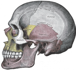
Sutures are fibrous joints found exclusively in the skull. These bones are bound together by Sharpey's fibres. While they are generally considered immovable, a tiny amount of movement is permitted, which provides the skull with necessary compliance and elasticity.
In fetal skulls, sutures are wide to facilitate movement during birth. The resulting "soft spots" are known as fontanelles. If these bones fuse prematurely before birth, the condition is called craniosynostosis. In adulthood, these joints may eventually ossify, turning completely into bone.
Syndesmosis
A syndesmosis is a slightly movable (amphiarthrodial) joint where bones are joined by a ligament or an interosseous membrane. Common examples include the connection between the radius and ulna in the forearm and the distal tibiofibular joint in the ankle.
Gomphosis
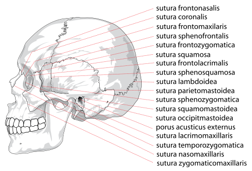
A gomphosis, often described as a "peg and socket joint," binds the root of a tooth to the socket of the maxilla (upper jaw) or mandible (lower jaw). The connection is maintained by the periodontal ligament, which attaches the jawbone to the cementum of the tooth.
Detailed Analysis of Cranial Sutures
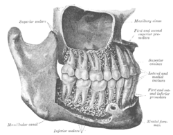
Cranial sutures are categorized by the shape of the interlocking bone edges:
- Serrate: Interlocking regions are serrated (e.g., Coronal and Sagittal sutures).
- Plane: Edges are flush, similar to a butt joint (e.g., Internasal suture).
- Limbous: Bevelled edges creating a sloping plane (e.g., Temporo-parietal suture).
- Schindylesis: Two bones fitting together like a bridle joint (e.g., Palatomaxillary suture).
- Denticulate: Edges that slot together like a finger joint (e.g., Lambdoid suture).
Major Named Sutures
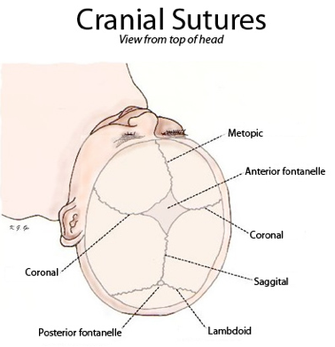
While many sutures are named after the bones they connect, several have distinct names based on their location:
- Coronal Suture: Between the frontal and parietal bones.
- Sagittal Suture: Along the midline between the parietal bones.
- Lambdoid Suture: Between the parietal and occipital bones.
- Squamosal Suture: Between the parietal and temporal bones.
Clinical Significance: Syndesmotic Injuries
Injuries to the distal tibiofibular syndesmosis are known as high ankle sprains. These are diagnosed through physical examinations using the squeeze test and the external rotation test. High-grade injuries often prevent a patient from performing a single-leg heel raise.
Grading Syndesmotic Tears
| Grade | Injury Description | Clinical Findings | Treatment Approach |
|---|---|---|---|
| Grade I | Partial anteroinferior tibiofibular ligament tear | Negative squeeze and exorotation tests | Conservative management |
| Grade II | Complete anteroinferior tibiofibular and inferior interosseous ligament tear | Positive squeeze and exorotation tests | Immobilization |
| Grade III | Complete anteroinferior tibiofibular tear, partial interosseous tear, and deltoid ligament avulsion | Positive squeeze and exorotation tests; unstable joint | Operative stabilization |
Surgical stabilization may involve a syndesmotic screw, which temporarily inhibits movement until healing occurs, or a tightrope fixation, which uses elastic fiberwire to allow physiologic motion.
Frequently Asked Questions
What is the difference between a suture and a syndesmosis?
A suture is an immovable (synarthrodial) joint found only in the skull, whereas a syndesmosis is a slightly movable (amphiarthrodial) joint found in other parts of the body, such as the forearm and ankle.
Why are fontanelles important in infants?
Fontanelles are the "soft spots" created by unfused cranial sutures. They are critical because they allow the skull to deform slightly during birth and provide room for the brain to grow during early development.
Is a tooth considered a bone in a gomphosis joint?
No, teeth are not technically bone. This makes the gomphosis unique, as it is the only fibrous joint where a bone (the jaw) does not join another bone.
How do orthodontists move teeth if gomphoses are fixed joints?
Although the motion of a gomphosis is minimal, it is not absolute. Considerable movement can be achieved over time through the application of constant pressure, which is the mechanical basis for using braces to realign teeth.
What is the difference between a screw and a tightrope fixation for ankle injuries?
A syndesmotic screw provides rigid fixation that inhibits normal bone movement and is usually removed after healing. A tightrope fixation uses elastic fiberwire to allow natural physiologic motion and may be left in place permanently.