Mitosis: The Process of Eukaryotic Cell Division
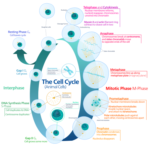
In the complex world of eukaryotic cells, growth and repair depend on a precise mechanism of duplication known as mitosis. Mitosis is a specialized part of the cell cycle where replicated chromosomes are separated into two new nuclei. This process is an equational division, meaning it produces two genetically identical daughter cells that maintain the same total number of chromosomes as the original mother cell.
To ensure genetic stability across generations, mitosis is carefully timed. It is preceded by the S phase of interphase, during which DNA replication occurs. Following the nuclear division, the cell undergoes telophase and cytokinesis to divide the cytoplasm, organelles, and cell membrane. Together, these stages define the mitotic phase (M phase).
Key Facts
- Genetic Identity: Mitosis results in two daughter cells that are genetically identical to the parent cell.
- Chromosome Maintenance: The total number of chromosomes is preserved throughout the division.
- Prerequisite: DNA must be replicated during the S phase of interphase before mitosis can begin.
- Outcome: The process concludes with cytokinesis, splitting one cell into two.
The Stages of Mitosis
Mitosis is divided into distinct stages based on the cellular activities occurring at each step. While the general process is consistent, some variations exist, such as the preprophase stage found specifically in plant cells.
Prophase and Prometaphase
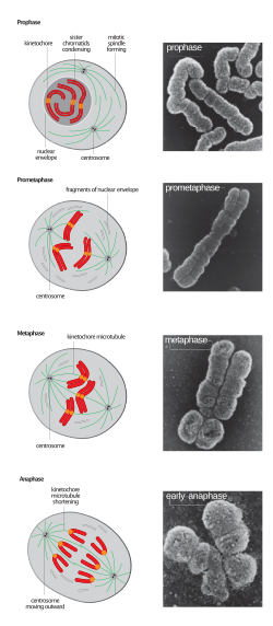
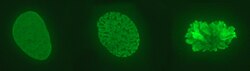
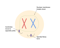
During prophase, the duplicated chromosomes condense, making them visible under a microscope. They attach to spindle fibers, which are protein structures that will eventually pull the genetic material apart. This is followed by prometaphase, where the nuclear envelope breaks down, allowing the spindle fibers to access the chromosomes.
Metaphase
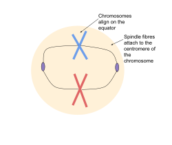
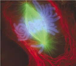
In metaphase, the spindle fibers align the chromosomes along the center of the cell, known as the metaphase plate. This alignment ensures that when the chromosomes separate, each new nucleus will receive exactly one copy of each chromosome.
Anaphase
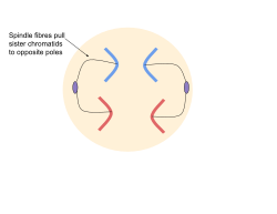
During anaphase, the sister chromatids (the two identical halves of a duplicated chromosome) are pulled apart toward opposite poles of the cell. This movement is driven by the shortening of the spindle fibers.
Telophase and Cytokinesis
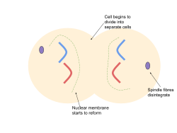
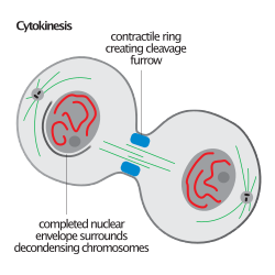
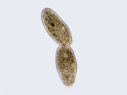
Telophase is the final stage of nuclear division, where two new nuclei form around the separated sets of chromosomes. However, the cell is not yet fully divided. Cytokinesis follows, dividing the cytoplasm and cell membrane to create two independent daughter cells.
Cellular Dynamics and Variations
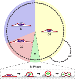
Mitosis involves significant physical changes to the cell. In many animal cells, mitotic cell rounding occurs; the cell changes shape to facilitate the assembly of the spindle. This process involves the actomyosin cortex and is essential for efficient division.
Forms and Errors
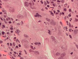
Depending on the organism, mitosis can take different forms, such as open orthomitosis or closed intranuclear pleuromitosis. When mitosis fails, errors can occur. For example, tripolar mitosis is an abnormal division that can be seen in precancerous lesions, such as those in the stomach or breast cancer tissues.
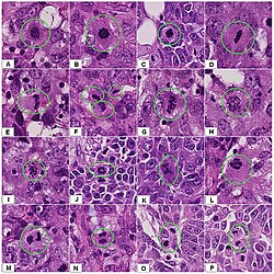
Summary of Mitotic Process
| Stage | Primary Action | Key Outcome |
|---|---|---|
| Prophase | Chromosome condensation | Visible chromosomes and spindle formation |
| Metaphase | Alignment at the center | Chromosomes positioned at the metaphase plate |
| Anaphase | Separation of chromatids | Identical genetic sets move to opposite poles |
| Telophase | Nuclear envelope reformation | Two distinct daughter nuclei |
| Cytokinesis | Cytoplasmic division | Two separate, genetically identical cells |
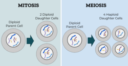
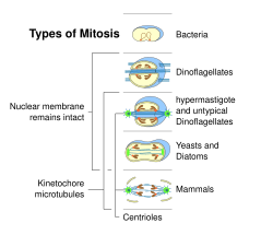
Frequently Asked Questions
How does mitosis differ from meiosis?
Mitosis produces two genetically identical daughter cells for growth and repair, whereas meiosis is used to produce gametes (sex cells) with half the original number of chromosomes.
What happens during the S phase before mitosis?
The S phase is the period of interphase where DNA replication occurs, ensuring that there are two complete sets of chromosomes available to be divided during mitosis.
What is the role of the spindle fibers?
Spindle fibers attach to the chromosomes and physically pull the sister chromatids apart toward opposite ends of the cell during anaphase.
What is cytokinesis?
Cytokinesis is the physical process of dividing the cell's cytoplasm, organelles, and membrane, which officially separates the mother cell into two distinct daughter cells.
Can mitosis go wrong?
Yes. Errors such as tripolar mitosis can occur, where chromosomes are pulled in three directions instead of two. Such abnormalities are often associated with precancerous lesions and cancer.