Karyotypes: The Science of Chromosome Analysis
In the realm of genetics, a karyotype represents the complete set of chromosomes within the cells of a species or a specific individual. By examining the size, number, and shape of these structures, scientists can gain critical insights into the biological blueprint of an organism. The process of determining this complement is known as karyotyping, a diagnostic and research tool used to identify chromosomal abnormalities and understand evolutionary relationships.
When a karyotype is visualized as a graphical depiction, it is called a karyogram (or idiogram). In these charts, chromosomes are typically organized in pairs and ordered by size and the position of the centromere—the constricted region that joins sister chromatids.

Key Facts
- Human Count: A normal diploid human cell contains 46 chromosomes (23 pairs).
- Karyogram Types: Micrographic karyograms use actual photographs of cells, while schematic karyograms are designed graphic representations.
- Cell Cycle Timing: Karyotyping is generally performed during metaphase, when chromosomes are most condensed and visible.
- Fundamental Number (FN): This refers to the number of visible major chromosomal arms per set; for humans, FN = 82.
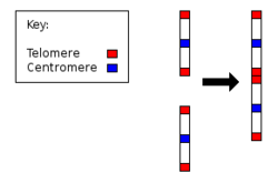
- Evolutionary Marker: Human chromosome 2 resulted from the fusion of two ancestral ape chromosomes, reducing the human count to 46 compared to the 48 found in great apes.
The Process of Karyotyping
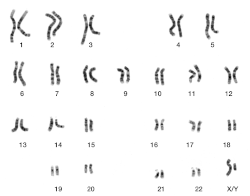
Karyotyping combines light microscopy and photography to capture the genome. To produce a micrographic karyogram, cells are typically processed through several steps: they are grown in tissue culture, treated with a hypotonic solution to swell the cells and spread the chromosomes, and arrested in metaphase using colchicine. Finally, the preparation is squashed on a slide to force the chromosomes into a single plane for clear imaging.
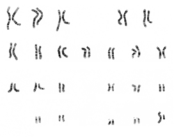
While chromosomes naturally appear purple when stained with Giemsa, micrographs are often converted to grayscale to make comparisons between different laboratories easier.
![Micrograph of human chromosomes before further processing. Staining with Giemsa confers a purple color to chromosomes, but micrographs are often converted to grayscale to facilitate data presentation and make comparisons of results from different laboratories.[7]](/images/8a/9e/8a9ea77da4bdea568dba11bd02a0e1a35e7d667c63327cbcf7d4f10d76d8cf2d.jpg)
Understanding Copy Number and the Cell Cycle
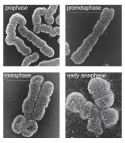
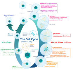
The appearance of chromosomes changes depending on the phase of the cell cycle. In the G0 and G1 phases, DNA exists as dispersed chromatin and is not visually distinguishable. During the S phase, DNA synthesis occurs. By the G2 phase and metaphase, each chromosome consists of two identical sister chromatids connected at the centromere. Although the chromosome count (2n) remains the same, the DNA content doubles (from 2c to 4c).
Human Chromosome Classification
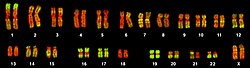
Human chromosomes are categorized into groups based on their size and the position of the centromere. Centromere position defines whether a chromosome is metacentric (centromere in the middle), submetacentric (centromere slightly off-center), or acrocentric (centromere near one end, often with a satellite).
| Group | Chromosomes | Physical Features |
|---|---|---|
| A | 1–3 | Large, metacentric or submetacentric |
| B | 4–5 | Large, submetacentric |
| C | 6–12, X | Medium-sized, submetacentric |
| D | 13–15 | Medium-sized, acrocentric, with satellite |
| E | 16–18 | Small, metacentric or submetacentric |
| F | 19–20 | Very small, metacentric |
| G | 21–22, Y | Very small, acrocentric (21, 22 with satellite) |
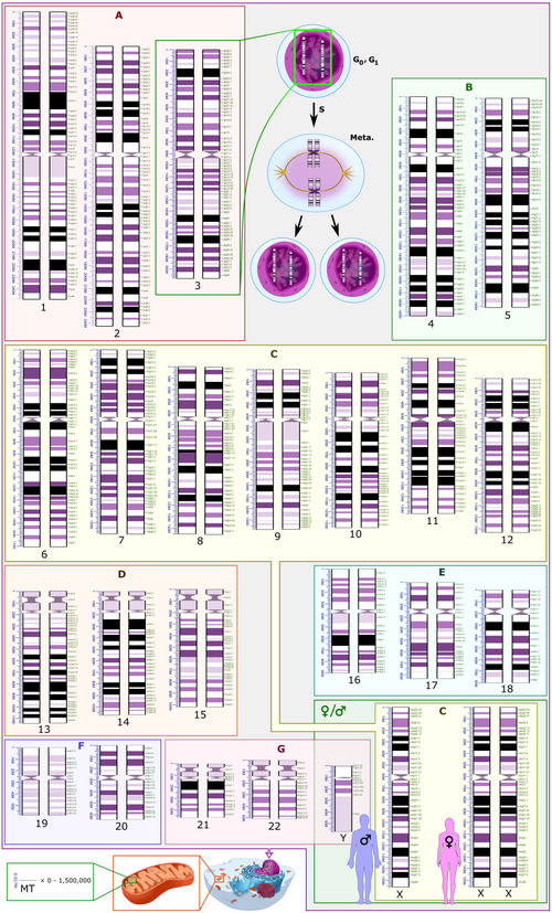
Advanced Visualization Techniques
Modern cytogenetics uses various banding and staining techniques to identify specific regions of chromosomes. The short arm is designated as p and the long arm as q. Numerical designations are assigned from the proximal (center) to the distal (end) regions of the arms.
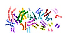
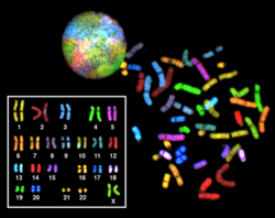
Beyond classic staining, scientists use Fluorescence In Situ Hybridization (FISH) to probe for specific DNA sequences, and Spectral Karyotyping (SKY) or multicolor FISH (mFISH) to paint each chromosome pair a different color, allowing for the rapid detection of translocations and other abnormalities.
Chromosomal Diversity and Abnormalities
Variation in karyotypes can occur between sexes, between germ-line and soma cells, or among members of a population (chromosome polymorphism). In some cases, aneuploidy—an abnormal number of chromosomes—occurs. This is seen in various plant genera like Crepis and Crocus, where haploid numbers vary significantly between species.
In humans, chromosomal abnormalities can lead to disease. For example, Cri du chat syndrome is caused by a deletion on the short arm of chromosome 5 (specifically region p15.2), denoted in cytogenetic notation as 46,XX,del(5)(p15.2).
Frequently Asked Questions
What is the difference between a karyotype and a karyogram?
A karyotype is the actual set of chromosomes in a cell, while a karyogram is the visual image or chart where those chromosomes are organized and paired for analysis.
Why are chromosomes photographed during metaphase?
During metaphase, chromosomes are at their most condensed state, making them clearly visible and distinguishable under a light microscope.
How many chromosomes do humans have compared to other great apes?
Humans have 46 chromosomes (23 pairs), whereas other great apes have 48. This difference is due to the fusion of two ancestral chromosomes to form human chromosome 2.
What does the term "acrocentric" mean?
An acrocentric chromosome is one where the centromere is located very close to one end, resulting in one very short arm and one long arm, often featuring a satellite.
What is the purpose of FISH in karyotyping?
Fluorescence In Situ Hybridization (FISH) uses fluorescent probes to bind to specific DNA sequences, allowing researchers to locate specific genes or detect deletions and duplications that are too small to see with standard staining.