Lungs: Anatomy, Function, and Biological Development
The lungs are the primary organs of the respiratory system, serving as the critical interface where the body interacts with the atmosphere to sustain life. Situated within the thoracic cavity of the chest, these paired organs flank the heart and great vessels, working tirelessly to ensure that oxygen enters the bloodstream and carbon dioxide is removed.
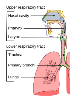
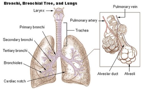
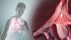
From a structural perspective, the lungs are part of the lower respiratory tract. This system begins at the trachea, which branches into the bronchi and further into smaller bronchioles. This network, known as the conducting zone, directs air toward the microscopic alveoli, where the essential process of gas exchange occurs.
Key Facts
- Total Weight: Approximately 1.3 kilograms (2.9 lb) combined.
- Airway Length: The lungs contain roughly 2,400 kilometers (1,500 mi) of airways.
- Alveoli Count: Between 300 to 500 million microscopic air sacs.
- Surface Area: Gas exchange occurs across a massive surface area estimated between 70 and 145 square meters.
- Lobe Distribution: The right lung has three lobes, while the left lung has two to accommodate the heart.
Anatomical Structure and Organization
Humans possess two lungs: a right and a left. The right lung is generally larger and heavier than the left. To protect these organs and facilitate movement, each lung is enclosed in a pleural sac consisting of two pleurae. This arrangement allows the inner and outer walls to slide against each other with minimal friction during the act of breathing.
Lobes and Fissures
The inner visceral pleura divides the lungs into sections called lobes via fissures. The right lung is divided into three lobes (Upper, Middle, and Lower), while the left lung contains only two (Upper and Lower). These lobes are further subdivided into bronchopulmonary segments and lobules.
It is important to note that anatomical variations are common. For instance, a horizontal interlobar fissure may be incomplete in 25% of right lungs or entirely absent in 11% of cases. Some individuals may even have a right lung with only two lobes or a left lung with three.
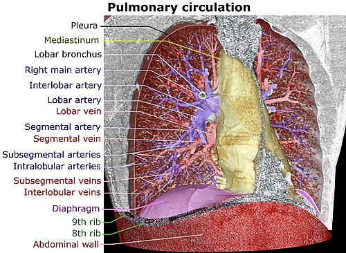
Microanatomy and the Respiratory Zone
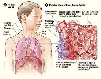
At the microscopic level, the lungs are a complex network of connective tissue and respiratory epithelium. The terminal bronchioles branch into respiratory bronchioles, which supply the alveoli held within each acinus.
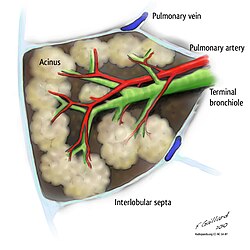
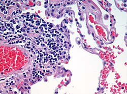
The alveoli are the functional units of the lung. These tiny sacs are surrounded by dense capillary networks, creating a thin blood-air barrier (approximately 0.5 to 2 μm thick) that allows gases to equilibrate rapidly between the air and the blood.
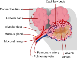
Blood Supply and Circulation
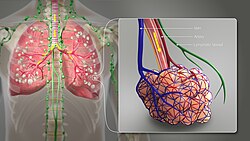
The lungs feature a unique dual blood supply system to meet two different needs:
- Pulmonary Circulation: Deoxygenated blood is sent from the heart to the lungs to be oxygenated.
- Bronchial Circulation: A separate supply of oxygenated blood that provides nutrients to the lung tissue itself.
Biological Development
The development of the lungs follows a process called branching morphogenesis, where the respiratory tree is formed through the repeated splitting of bronchial buds. This process is governed by specific genes and signaling proteins, most notably FGF10 (fibroblast growth factor 10), which is essential for epithelial branching. Other contributing factors include sonic hedgehog (SHH) and bone morphogenetic protein BMP4.
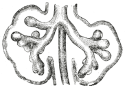
While the bronchi and bronchioles form through bifurcation, the development of the alveoli occurs when this branching stops and the distal tips dilate to form the air sacs.
Comparative Biology: Lungs in Other Animals
While human lungs are specialized for mammalian needs, other species have evolved diverse respiratory strategies:
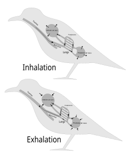
- Birds: Utilize a unidirectional flow system with air sacs that force air through parabronchi, employing a cross-current gas exchanger for high efficiency.
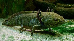
- Amphibians: Some, like the axolotl, retain larval gills into adulthood.
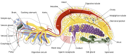
- Invertebrates: Spiders utilize "book lungs," which are stacks of hemolymph-filled lamellae.
![The cross-current respiratory gas exchanger in the lungs of birds. Air is forced from the air sacs unidirectionally (from left to right in the diagram) through the parabronchi. The pulmonary capillaries surround the parabronchi in the manner shown (blood flowing from below the parabronchus to above it in the diagram).[103][104] Blood or air with a high oxygen content is shown in red; oxygen-poor air or blood is shown in various shades of purple-blue.](/images/2c/0e/2c0e18e79fd622b616d7fcf53153175511234a11334c7adb640cf1cace938998.jpg)
Summary of Lung Characteristics
| Feature | Right Lung | Left Lung |
|---|---|---|
| Number of Lobes | 3 (Upper, Middle, Lower) | 2 (Upper, Lower) |
| Relative Size | Larger and Heavier | Smaller (shares space with heart) |
| Weight (Men) | 155–720 g | 110–675 g |
| Weight (Women) | 100–590 g | 105–515 g |
Frequently Asked Questions

What is the primary function of the lungs?
The primary function is gas exchange. The lungs bring oxygen from the air into the blood and remove carbon dioxide from the blood to be exhaled.
Why is the left lung smaller than the right lung?
The left lung is smaller because it must share space within the thoracic cavity with the heart.
What are alveoli and why are they important?
Alveoli are microscopic air sacs where the actual exchange of oxygen and carbon dioxide takes place. Their vast number and thin walls provide the necessary surface area for efficient diffusion.
What is branching morphogenesis?
It is the developmental process by which the lungs form their tree-like structure through the repeated splitting and growth of bronchial buds, largely driven by the protein FGF10.
How does the blood supply to the lungs work?
The lungs have two systems: the pulmonary circulation, which brings deoxygenated blood to be refreshed with oxygen, and the bronchial circulation, which provides oxygenated blood to the lung's own tissues.