Respiratory System: Anatomy, Gas Exchange, and Comparative Biology
The respiratory system is a complex biological network designed to facilitate the exchange of gases between an organism and its environment. In humans and other mammals, this system ensures that oxygen is absorbed into the bloodstream to fuel cellular metabolism, while carbon dioxide—a waste product of these processes—is efficiently removed.
From the initial intake of air to the microscopic diffusion of gases in the lungs, the process involves a sophisticated series of anatomical structures and physiological mechanisms that adapt to varying environmental pressures, including high altitudes and aquatic habitats.
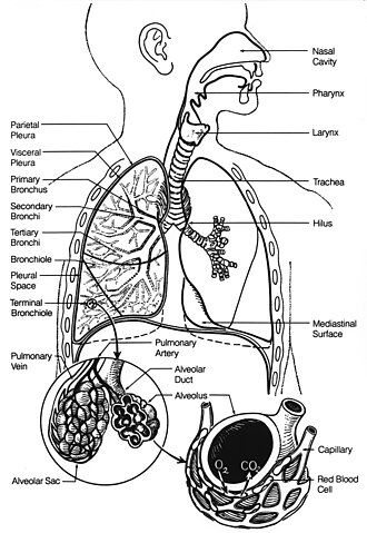
Mammalian Respiratory Anatomy
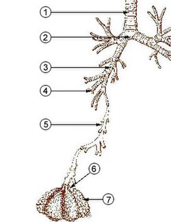
The human respiratory tract is divided into upper and lower sections. The lower tract is often visualized as a respiratory tree (or tracheobronchial tree), characterized by a series of branching generations. In an adult human, there are approximately 23 generations of these branches.
The Conducting Zone
Generations 0 through 16 serve primarily as air conduits. This zone begins with the trachea, which branches into the right and left main bronchi. These main bronchi enter the lungs at the hilum and further divide into secondary (lobar) bronchi and tertiary (segmental) bronchi. As the pathways narrow, they become subsegmental bronchi and eventually bronchioles—small airways that lack cartilaginous support.
The Respiratory Zone
Gas exchange occurs in the final generations (17–23), which include the respiratory bronchioles, alveolar ducts, and alveoli. Alveoli are tiny air sacs, ranging from 75 to 300 μm in diameter. With roughly 300 million alveoli, the lungs provide a massive surface area of approximately 145 m² for efficient gas diffusion.
Mechanics of Breathing and Ventilation
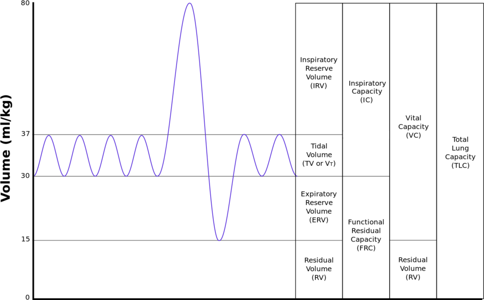
Breathing is the physical process of moving air into and out of the lungs. The volume of air displaced during a single cycle is known as the tidal volume, which averages 500 ml in a resting adult.
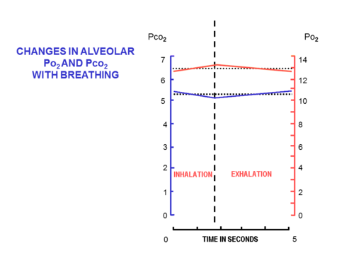
Not all inhaled air reaches the gas-exchange surfaces. About 150 ml remains in the airways, a phenomenon known as dead space ventilation. Consequently, only about 350 ml of fresh, moistened air reaches the alveoli with each breath. This fresh air mixes with the functional residual capacity (2.5–3.0 liters of air remaining after exhalation), ensuring that the composition of alveolar air remains relatively stable throughout the breathing cycle.
The partial pressures of gases in the alveoli are significantly different from those in dry outside air at sea level. While atmospheric oxygen partial pressure is 21 kPa, alveolar oxygen remains around 13–14 kPa. Carbon dioxide in the alveoli is maintained at approximately 5.3 kPa, compared to 0.04 kPa in the ambient air.
The Process of Gas Exchange
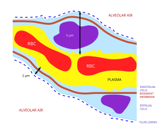
The primary goal of the respiratory system is to equalize the partial pressures of gases between the alveolar air and the pulmonary capillary blood. This occurs via simple diffusion across the blood-air barrier, an extremely thin membrane (averaging 2.2 μm) composed of alveolar epithelial cells, basement membranes, and capillary endothelial cells.
Oxygen and Carbon Dioxide Transport
- Oxygen: Because oxygen has low solubility in water, it is transported by combining with hemoglobin. Each hemoglobin molecule contains four iron-containing heme groups. When all four are occupied, the blood is considered "saturated."
- Carbon Dioxide: Most CO² is transported as bicarbonate ions (HCO³-) in the plasma. This conversion is accelerated by the enzyme carbonic anhydrase found within red blood cells. A small portion is also carried as carbamino groups on hemoglobin.
Adaptations to High Altitude
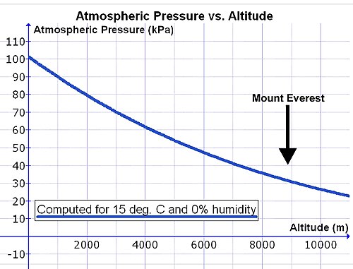
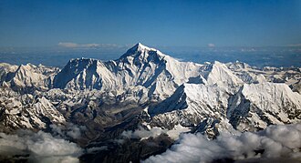
At high altitudes, the total atmospheric pressure drops, which reduces the partial pressure of oxygen. For example, at the summit of Mount Everest, the total pressure is only 33.7 kPa. Because the body must still saturate inhaled air with water vapor (6.3 kPa at 37°C), the available oxygen pressure entering the alveoli drops to 5.8 kPa.
To compensate, the body employs hyperpnea (deeper and faster breathing). At sea level, the body prioritizes the regulation of carbon dioxide levels. However, above 2,500 meters, the system switches priority to oxygen homeostasis. This shift can cause a severe drop in arterial CO² and a rise in plasma pH, contributing to high altitude sickness.
Comparative Respiratory Systems
Avian Respiration
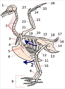
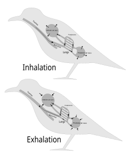
Birds possess a highly efficient unidirectional flow system. Unlike mammals, they utilize air sacs that act as bellows to push air through the lungs (parabronchi) during both inhalation and exhalation.
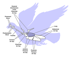
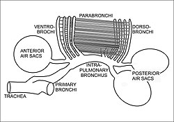
![Fig. 19 The cross-current respiratory gas exchanger in the lungs of birds. Air is forced from the air sacs unidirectionally (from right to left in the diagram) through the parabronchi. The pulmonary capillaries surround the parabronchi in the manner shown (blood flowing from below the parabronchus to above it in the diagram).[45][48] Blood or air with a high oxygen content is shown in red; oxygen-poor air or blood is shown in various shades of purple-blue.](/images/2c/0e/2c0e18e79fd622b616d7fcf53153175511234a11334c7adb640cf1cace938998.jpg)
Aquatic Respiration (Fish)
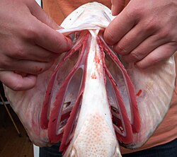
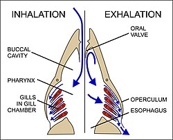
Oxygen is far less soluble in water than in air, and diffuses 10,000 times slower. Fish overcome this using gills, which contain filaments and lamellae. They utilize a countercurrent flow exchange system, where blood flows in the opposite direction to the water, maximizing oxygen extraction from the environment.
![Fig. 22. A comparison between the operations and effects of a cocurrent and a countercurrent flow exchange system is depicted by the upper and lower diagrams respectively. In both, it is assumed that red has a higher value (e.g. of temperature or the partial pressure of a gas) than blue and that the property being transported in the channels, therefore, flows from red to blue. In fish a countercurrent flow (lower diagram) of blood and water in the gills is used to extract oxygen from the environment.[55][56][57]](/images/f0/3e/f03e21c35c56e9fdf3bf8c3445c665c337a9f62a7ffba1f140ec1c2566c4502b.webp)
Other Organisms
- Cetaceans: Marine mammals can replace up to 90% of their lung volume per breath and store extra oxygen in muscles via myoglobin.
- Plants: Respiration occurs via stomata (small pores). While their oxygen needs are low, their demand for CO² for photosynthesis is very high.
Key Facts
- Alveolar Surface Area: The human lungs provide approximately 145 m² for gas exchange.
- Blood-Air Barrier: The membrane through which gases diffuse is only about 2.2 μm thick.
- Tidal Volume: A resting adult breathes roughly 500 ml of air per cycle.
- Altitude Switch: At approximately 2,500 m, the body prioritizes oxygen homeostasis over carbon dioxide regulation.
- Avian Efficiency: Birds use a cross-current exchange system with unidirectional airflow.
- Fish Strategy: Countercurrent flow in gills allows fish to extract oxygen from water despite low solubility.
| Measurement | Equation | Description |
|---|---|---|
| Minute ventilation | Tidal volume × Respiratory rate | Total volume of air breathed per minute |
| Alveolar ventilation | (Tidal volume − Dead space) × Respiratory rate | Volume of fresh air reaching the alveoli per minute |
| Dead space ventilation | Dead space × Respiratory rate | Volume of air that does not participate in gas exchange |
Clinical Significance
Respiratory disorders are generally categorized into several groups:
- Obstructive: Emphysema, bronchitis, and asthma.
- Restrictive: Fibrosis, sarcoidosis, and pleural effusion.
- Vascular: Pulmonary edema, embolism, and hypertension.
- Infectious/Environmental: Pneumonia, tuberculosis, and asbestosis.
- Other: Primary and secondary cancers, and respiratory distress syndrome (insufficient surfactant) in pre-term infants.
Frequently Asked Questions
What is the difference between tidal volume and alveolar ventilation?
Tidal volume is the total amount of air moved in or out of the lungs during a single breath (about 500 ml). Alveolar ventilation is the portion of that air that actually reaches the alveoli for gas exchange, excluding the air that remains in the "dead space" of the conducting airways.
How does the body adapt to breathing at high altitudes?
The body engages in hyperpnea, breathing deeper and faster to compensate for the lower partial pressure of oxygen. Above 2,500 meters, the body shifts its homeostatic priority from regulating carbon dioxide to regulating oxygen.
Why is the countercurrent system in fish gills important?
Because oxygen diffuses much slower in water than in air and is present in lower concentrations, the countercurrent system (where blood and water flow in opposite directions) ensures a constant concentration gradient, allowing fish to extract the maximum amount of oxygen possible.
What role does hemoglobin play in the respiratory system?
Hemoglobin is a protein in red blood cells that binds to oxygen via four iron-containing heme groups. This allows the blood to carry significantly more oxygen than could be dissolved in the plasma alone.
What is the blood-air barrier?
The blood-air barrier is the extremely thin membrane (approx. 2.2 μm) consisting of alveolar epithelial cells and capillary endothelial cells. It is the physical interface where oxygen diffuses into the blood and carbon dioxide diffuses into the alveoli.