Circulatory System: Anatomy, Function, and Evolutionary History
The circulatory system is the body's sophisticated transport network, responsible for delivering oxygen, nutrients, and essential chemicals to cells while removing metabolic waste. In humans, this system is a closed loop powered by the heart, ensuring that blood reaches every corner of the body to maintain homeostasis.
An average adult carries approximately five to six quarts (4.7 to 5.7 liters) of blood, which makes up about 7% of total body weight. This vital fluid is composed of plasma, red blood cells, white blood cells, and platelets. To function effectively, the circulatory system works in tandem with the digestive system to acquire the nutrients necessary to keep the heart pumping.
Key Facts
- Dual Circuits: The system is divided into pulmonary (lungs) and systemic (body) circulations.
- Oxygen Transport: Approximately 98.5% of arterial oxygen is bound to hemoglobin.
- Vessel Scale: Muscle capillaries in a 70 kg human can stretch between 9,000 and 19,000 km.
- Blood Volume: Blood accounts for roughly 7% of an adult's total body weight.
- Development: Fetal circulation bypasses the lungs via the truncus arteriosus.
The Two Major Circuits
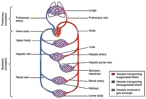
The human circulatory system is organized into two primary loops that ensure blood is oxygenated before being distributed to the body's tissues.
Pulmonary Circulation
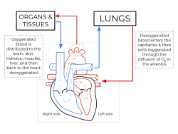
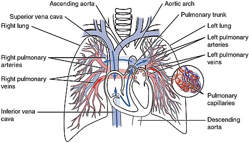
The pulmonary circulation is the loop that carries deoxygenated blood from the right side of the heart to the lungs. Once in the lungs, the blood is oxygenated and then returns to the left side of the heart.
Systemic Circulation
The systemic circulation delivers oxygen-rich blood from the left heart to the rest of the body. After delivering oxygen to the tissues, the deoxygenated blood returns to the right heart through large veins called the venae cavae. This circuit is further categorized into macrocirculation (large vessels) and microcirculation (small vessels).
Blood Vessels and Transport
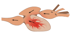
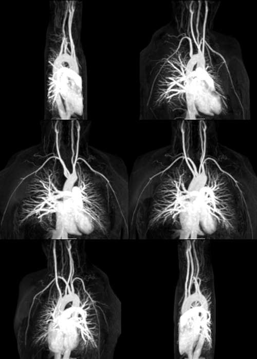
Blood travels through a network of vessels categorized by their structure and the direction of blood flow. The largest arteries and veins connecting directly to the heart are known as the great vessels.
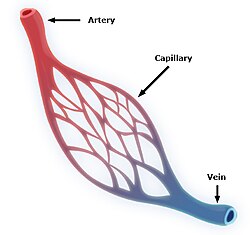
- Arteries: These carry blood away from the heart. They branch into smaller passages called arterioles.
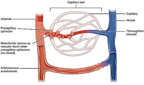
- Capillaries: These are microscopic vessels that join the arterial and venous systems. They are the primary site of exchange between blood and tissues.
- Veins: These return blood to the heart.
The efficiency of this system is highlighted by the renal circulation, which supplies the kidneys. This specialized network receives approximately 20% of the total cardiac output, branching from the abdominal aorta and returning via the ascending inferior vena cava.
Development and Function
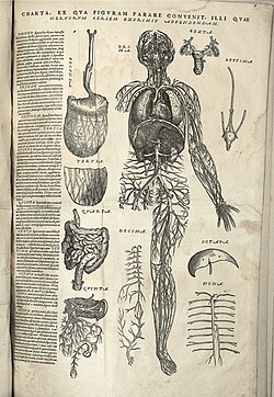
The circulatory system begins forming in the embryo through a process called vasculogenesis. The arterial system develops primarily from six pairs of aortic arches, while the venous system arises from three bilateral veins between weeks 4 and 8 of embryogenesis.
Fetal circulation is distinct because it bypasses the lungs, as the fetus obtains oxygen and nutrients from the mother via the placenta and umbilical cord. This bypass occurs through the truncus arteriosus until birth.
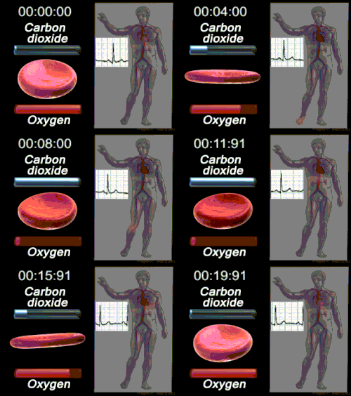
In a healthy adult at sea level, the primary transporter of oxygen is the hemoglobin molecule. About 98.5% of arterial oxygen is chemically combined with hemoglobin, while only 1.5% is physically dissolved in the blood plasma.
Clinical Significance and Treatment
Diseases affecting this system are collectively known as cardiovascular disease. Modern medicine employs several surgical interventions to treat these conditions:
- Angioplasty: A procedure to open narrowed blood vessels.
- Coronary Artery Bypass/Stents: Used to restore blood flow to the heart muscle.
- Valve Repair: Treatment for damaged heart valves.
- Phlebectomy: The removal of varicose veins.
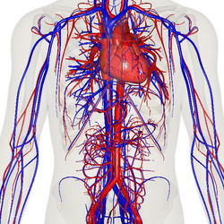
In the United States, the majority of these procedures are performed in inpatient settings, with only 28% occurring in ambulatory care settings.
Comparative Anatomy and Evolution
The vascular system evolved over millions of years to overcome the limitations of diffusion. The first blood vascular systems likely appeared over 600 million years ago, with endothelium evolving in ancestral vertebrates between 510 and 540 million years ago.
| System Type | Characteristics | Example |
|---|---|---|
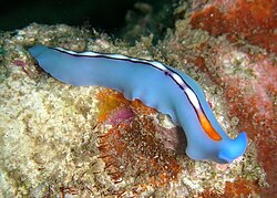
| No Circulatory System | Lack specialized circulatory organs | Flatworms (e.g., Pseudoceros bifurcus) |
| Open Circulatory System | Hemolymph pumped into a hemocoel | Grasshopper |
| Closed Circulatory System | Blood contained within vessels | Humans, Fish (two-chambered heart) |
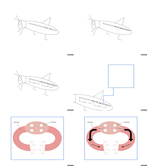
Historical Discoveries
Our understanding of the heart has evolved from ancient mysticism to precise science. The Ebers Papyrus (16th century BCE) first acknowledged a connection between the heart and arteries, though Egyptians believed arteries transported air.
Significant milestones include:
- Avicenna (1025): Provided the first correct explanation of pulsation as alternate expansion and contraction.
- Ibn al-Nafis (1242): Described pulmonary circulation and predicted the existence of capillaries (pores) 400 years before their discovery.
- Michael Servetus (1546): The first European to describe pulmonary function, though his work was suppressed.
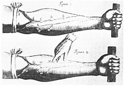
- William Harvey (1628): Demonstrated that the heart produces a continuous circulation of blood throughout the entire body.
- Marcello Malpighi (1661): Finally identified the capillary system connecting arteries and veins.
Frequently Asked Questions
What is the difference between pulmonary and systemic circulation?
Pulmonary circulation moves deoxygenated blood from the heart to the lungs to pick up oxygen, while systemic circulation delivers that oxygenated blood from the heart to the rest of the body's tissues.
How much blood does the average adult have?
An average adult contains between five to six quarts (approximately 4.7 to 5.7 liters) of blood, which represents about 7% of their total body weight.
What is the role of hemoglobin in the blood?
Hemoglobin is the primary transporter of oxygen in vertebrates; in healthy humans at sea level, it chemically combines with about 98.5% of the oxygen in arterial blood.
How does fetal circulation differ from adult circulation?
Fetal circulation bypasses the lungs via the truncus arteriosus because the fetus receives oxygen and nutrients from the mother through the placenta and umbilical cord rather than from breathing air.
Who discovered the continuous circulation of blood?
English physician William Harvey published the demonstration of continuous blood circulation in 1628, though the capillaries that connect the systems were later discovered by Marcello Malpighi in 1661.