Endocardium: Structure, Function, and Clinical Significance
The heart is a complex organ composed of several specialized layers, each serving a distinct purpose to ensure the efficient pumping of blood throughout the body. The endocardium is the innermost layer of tissue that lines the chambers of the heart. Biologically and embryologically, its cells are similar to the endothelial cells that line the body's blood vessels, providing a smooth, protective surface for the heart's interior.
To understand the endocardium, it is helpful to view it within the context of the entire heart wall. It sits beneath the myocardium—the thick, muscular tissue responsible for the heart's contractions. Outside of the myocardium lies the epicardium, the outermost layer. The entire organ is further encased in a fibrous sac called the pericardium, which contains a small amount of lubricating fluid.
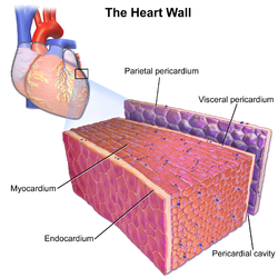
Key Facts
- Location: The innermost lining of the heart chambers and valves.
- Composition: Primarily composed of endothelial cells.
- Primary Role: Protects heart chambers and valves while modulating myocardial function.
- Barrier Function: Acts as a blood-heart barrier to regulate the ionic composition of extracellular fluid.
- Electrical Path: Depolarization impulses move from the endocardium to the epicardium.
The Vital Functions of the Endocardium
While it may seem like a simple lining, the endocardium plays a sophisticated role in controlling how the heart muscle functions. This modulating role is distinct from the homeometric and heterometric regulatory mechanisms that typically control myocardial contractility.
Modulating Heart Development and Function
The cardiac endothelium—which includes both the endocardial lining and the endothelium of the myocardial capillaries—is essential for heart development in embryos and adults. This is particularly evident during hypertrophy (the enlargement of cells). Furthermore, the endothelium helps regulate the electrophysiological environment and the contractility of cardiomyocytes (heart muscle cells).
The Blood-Heart Barrier
Similar to the blood-brain barrier, the endocardial endothelium functions as a blood-heart barrier. This allows the heart to precisely control the ionic composition of the extracellular fluid surrounding the cardiomyocytes, ensuring the electrical stability required for a steady heartbeat.
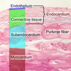
Clinical Significance and Pathology
Because of its position and function, the endocardium is central to several critical cardiac conditions. Understanding whether a pathology is subendocardial or transmural is vital for diagnosis and treatment.
Myocardial Infarction
In a myocardial infarction (heart attack), ischemia—the restriction of blood supply—typically begins at the endocardium. Depending on the severity, this can take two forms:
- Subendocardial Infarction: The damage is limited to the inner layer and does not reach the epicardium. In acute settings, these are particularly dangerous because they create dead tissue surrounded by damaged myocytes. This region conducts electrical impulses more slowly, which can lead to irregular heart rhythms.
- Transmural Infarction: The ischemia extends through the entire heart wall, from the endocardium to the epicardium. In chronic settings, these are more dangerous due to extensive muscular damage and the formation of scar tissue, which impairs the heart's ability to contract (systolic contractility) and relax (diastolic relaxation), increasing the risk of rupture and thrombus (blood clot) formation.
Infective Endocarditis
The endocardium is also the primary site of infective endocarditis, a condition where bacteria infect the endocardial lining, most commonly affecting the endocardium that lines the heart valves.
Summary of Heart Wall Layers
| Layer | Position | Primary Composition | Main Function |
|---|---|---|---|
| Endocardium | Innermost | Endothelial cells | Lining chambers/valves; modulating function |
| Myocardium | Middle | Muscular tissue | Contraction and pumping |
| Epicardium | Outermost | Connective tissue/Serosa | Protective outer covering |
| Pericardium | External Sac | Fibrous tissue | Enclosure and lubrication |
Frequently Asked Questions
What is the difference between the endocardium and the myocardium?
The endocardium is the thin, innermost lining of the heart chambers and valves, whereas the myocardium is the thick middle layer of muscle responsible for the heart's pumping action.
How does the endocardium act as a barrier?
The endocardial endothelium functions as a blood-heart barrier, regulating the ionic composition of the fluid surrounding the heart muscle cells, similar to how the blood-brain barrier protects the brain.
Why are subendocardial infarctions dangerous in acute settings?
They create an area of dead tissue surrounded by damaged myocytes that conduct electrical impulses slowly, which can trigger irregular and potentially life-threatening heart rhythms.
What is infective endocarditis?
Infective endocarditis is an infection caused by bacteria that attack the endocardium, specifically targeting the lining of the heart valves.
In which direction do electrical impulses travel during depolarization?
During depolarization, the electrical impulse is carried from the endocardium toward the epicardium.