Embryonic Cleavage: Mechanisms, Types, and Developmental Patterns
In the earliest stages of life, following fertilization but before the formation of the amnion, an embryo undergoes a critical process known as cleavage. This is a series of rapid cell divisions where the original zygote splits into numerous smaller cells. Unlike typical cell division, cleavage occurs without significant overall growth; the resulting cluster of cells remains the same size as the original zygote, meaning each new cell contains roughly half the cytoplasm of its parent.
These individual cells are called blastomeres. As they multiply, they form a compact, solid mass known as a morula (Latin for mulberry). This process eventually concludes with the formation of a hollow sphere called a blastula, or a blastocyst in the case of mammals.
Key Facts
- Cleavage increases cell number and nuclear mass without increasing total cytoplasmic volume.
- The type of cleavage is primarily determined by the concentration and distribution of yolk in the egg.
- Holoblastic cleavage is complete division, while meroblastic cleavage is partial.
- Determinate cleavage fixes cell fates early, whereas indeterminate cleavage allows cells to remain flexible.
- The midblastula transition marks the end of cleavage and the start of zygotic transcription in non-mammals.
Fundamental Laws of Cleavage
Early embryological studies established four general laws that govern how cells divide during this stage:
- Balfour's Law: In complete (holoblastic) cleavage, the rate of division is inversely related to the amount of yolk; higher yolk concentrations slow down the division of both the nucleus and cytoplasm.
- Sack's Law: Daughter cells are typically equal in size, and successive planes of division occur at right angles to one another.
- Hertwig's Law: The nucleus and mitotic spindle align with the longest axis of the active protoplasm, ensuring the cell cleaves across its longest dimension.
The Biological Mechanism
The speed of these cell cycles is maintained by high levels of regulatory proteins, specifically cyclins and cyclin-dependent kinases (CDKs). The complex formed by cyclin B and CDK1, known as the maturation promoting factor (MPF), is what triggers the entry into mitosis.
Cleavage ends when the embryo begins zygotic transcription (the process where the embryo's own DNA starts directing development). In non-mammals, this is called the midblastula transition, which is triggered when the ratio of nuclear material to cytoplasm reaches approximately 1:6.
Types of Cleavage
Determinate vs. Indeterminate Cleavage
Cleavage is categorized by whether the fate of the blastomeres is fixed early on. Determinate (mosaic) cleavage, common in most protostomes, means the developmental fate of each cell is set early; a single blastomere cannot develop into a complete embryo. Conversely, indeterminate (regulative) cleavage, characteristic of deuterostomes, allows separated early cells to each develop into a whole, independent organism.
Holoblastic (Complete) Cleavage
Holoblastic cleavage occurs when the entire egg is divided. This is further divided into several patterns:
- Bilateral: The zygote is bisected into mirror-image left and right halves.
- Radial: Spindle axes are parallel or perpendicular to the polar axis, common in echinoderms and vertebrates.
- Rotational: One daughter cell divides meridionally while the other divides equatorially, as seen in the nematode C. elegans and mammals.
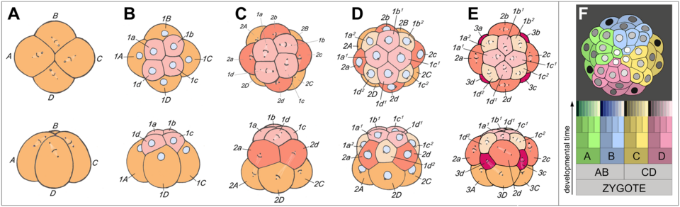
- Spiral: Common in lophotrochozoans (Spiralia), such as molluscs and annelids. Divisions occur at oblique angles, creating a spiral arrangement of cells when viewed from the animal pole.
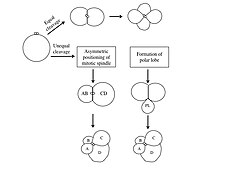
In spiral cleavage, the D macromere is vital for establishing the dorsal-ventral axis. This specification can happen through equal cleavage (where contact with micromeres determines the D quadrant) or unequal cleavage (via asymmetric spindles or a polar lobe).
Meroblastic (Incomplete) Cleavage
When an egg contains a high concentration of yolk, the cleavage furrows cannot penetrate the entire mass, resulting in meroblastic cleavage:
- Discoidal: The embryo forms a small disc of cells (blasto-disc) on top of the yolk. This is found in birds, reptiles, and most fish.
- Superficial: Mitosis occurs without cytokinesis, creating a multinucleated cell. Nuclei then migrate to the periphery to be partitioned into cells, a process common in arthropods like Drosophila.
Cleavage in Mammals
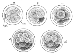
Mammalian cleavage is slower than in many other species, with divisions occurring every 12 to 24 hours. It is holoblastic and rotational. Zygotic transcription begins early, varying by species (e.g., at the two-cell stage in mice or the eight-cell stage in humans).
In humans, the embryo undergoes compaction at the eight-cell stage. Blastomeres flatten against each other, forming tight junctions and gap junctions. By the 16-cell stage, the embryo is a morula. The outer cells become trophoblasts, forming a watertight epithelial sheet enclosed within the zona pellucida. This structure eventually pumps in fluid to transform into a blastocyst.
Because of the indeterminate nature of early mammalian development, a single cell can be removed from a pre-compaction eight-cell embryo for genetic screening without harming the embryo's ability to develop.
Summary of Cleavage Types
| Cleavage Category | Yolk Distribution | Specific Pattern | Example Organisms |
|---|---|---|---|
| Holoblastic (Complete) | Isolecithal (Sparse/Even) | Radial | Echinoderms, Hemichordates |
| Spiral | Annelids, Most Mollusks | ||
| Bilateral | Tunicates | ||
| Rotational | Placental Mammals, Nematodes | ||
| Holoblastic (Displaced) | Mesolecithal (Moderate) | Displaced Radial | Amphibians, Lampreys |
| Meroblastic (Incomplete) | Telolecithal (Dense) | Discoidal / Bilateral | Birds, Reptiles, Cephalopods |
| Centrolecithal (Center) | Superficial | Most Insects |
Frequently Asked Questions
What is the difference between a morula and a blastocyst?
A morula is a solid, compact mass of blastomeres (typically around 16 cells) that resembles a mulberry. A blastocyst is the subsequent stage where the embryo develops a fluid-filled cavity and differentiates into an inner cell mass and an outer layer of trophoblasts.
How does yolk affect the way an embryo divides?
Yolk acts as a physical barrier to cleavage. In eggs with little yolk (isolecithal), the entire cell can divide (holoblastic). In eggs with dense yolk (telolecithal or centrolecithal), the cleavage furrows cannot pass through the yolk, leading to partial division (meroblastic).
What is the significance of the D macromere in spiral cleavage?
The D macromere is essential for establishing the secondary (dorsal-ventral) axis of the embryo. It directs the development of dorsal and posterior structures and signals to surrounding cells to determine their specific fates.
Why is indeterminate cleavage important for medical procedures?
Because cells in indeterminate cleavage retain the capacity to develop into a complete organism, a single blastomere can be removed from an early embryo (such as at the eight-cell stage in humans) for genetic screening without preventing the remaining cells from forming a healthy embryo.