Spinal Cord Anatomy, Function, and Clinical Significance
The spinal cord is a critical component of the central nervous system, serving as the primary communication highway between the brain and the peripheral nerves. Extending from the base of the skull through the vertebral column, it facilitates the transmission of sensory information to the brain and carries motor commands back to the muscles and organs.
In humans, the spinal cord begins at the occipital bone, exiting the skull through the foramen magnum and entering the spinal canal at the first cervical vertebra. While the vertebral column protects the cord, the spinal cord itself is shorter than the bony column it inhabits. In adult men, it typically measures around 45 cm (18 in), and in adult women, approximately 43 cm (17 in). Its diameter varies by region, ranging from 13 mm in the cervical and lumbar areas to 6.4 mm in the thoracic region.
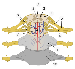
Key Facts
- Length: Approximately 43–45 cm in adults.
- Termination: Ends between the first and second lumbar vertebrae (L1–L2).
- Segments: Comprised of 31 distinct segments that give rise to pairs of spinal nerves.
- Protective Structure: Housed within the bony vertebral column.
- Key Features: Includes the conus medullaris (tapered end) and the cauda equina (bundle of nerve roots).
Structural Organization
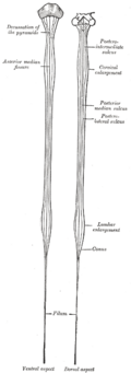
The spinal cord is ovoid in shape and features two distinct enlargements—areas where the cord widens to accommodate a higher density of neurons required for limb control.
Cervical and Lumbar Enlargements
- Cervical Enlargement: Stretching from C4 to T1, this region manages sensory and motor functions for the arms and trunk.
- Lumbar Enlargement: Located between T10 and L1, this region handles the sensory and motor input and output for the legs.
Segments and Spinal Nerves
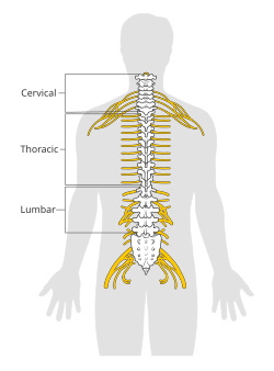
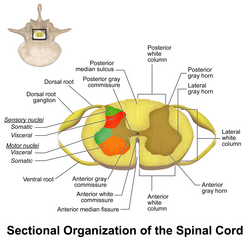
The cord is divided into 31 segments. Each segment produces one pair of sensory nerve roots (dorsal) and one pair of motor nerve roots (ventral). These roots merge to form mixed spinal nerves that exit the vertebral column through the intervertebral foramen.
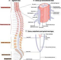
Interestingly, spinal cord segments do not align perfectly with the bony vertebrae in adults. Because the vertebral column grows faster than the spinal cord during development, the cord terminates at the L1–L2 level. This creates the conus medullaris, the tapered end of the cord. The nerves extending below this point to reach their respective exit holes form a bundle known as the cauda equina (Latin for "horse's tail").
| Species | Cervical | Thoracic | Lumbar | Sacral | Caudal/Coccygeal | Total |
|---|---|---|---|---|---|---|
| Human | 8 | 12 | 5 | 1 | - | 31 |
| Dog | 8 | 13 | 7 | 3 | 5 | 36 |
| Cat | 8 | 13 | 7 | 3 | 5 | 36 |
| Horse | 8 | 18 | 6 | 5 | - | 42 |
| Mouse | 8 | 13 | 6 | 4 | 3 | 35 |
Blood Supply and Development
The spinal cord relies on a complex network of arteries. Below the cervical region, the primary blood supply comes from posterior and anterior radicular arteries. A critical vessel in humans is the artery of Adamkiewicz (anterior radicularis magna), which typically arises between L1 and L2. Disruption of this artery, often during aortic surgery, can lead to spinal cord infarction and paraplegia.
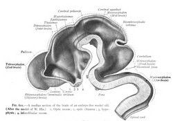
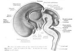
Neural Tracts and Motor Function
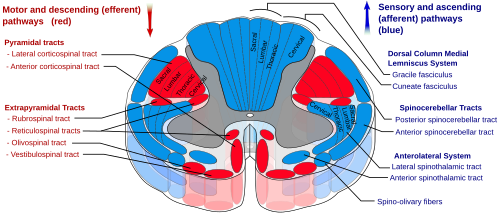
The spinal cord contains ascending tracts (which carry sensory data to the brain) and descending tracts (which carry motor commands to the muscles).
Ascending Tracts
Proprioceptive information (the sense of body position) travels via the dorsal spinocerebellar tract from L2 to T1, synapsing in Clarke's nucleus. Above T1, this information travels via the cuneocerebellar tract through the accessory cuneate nucleus before entering the cerebellum.
Descending Tracts and Motor Control
Motor commands originate in the cerebral cortex. About 90% of these axons cross over at the decussation of the pyramids in the medulla to form the lateral corticospinal tract, while 10% remain on the same side as the ventral corticospinal tract. These axons eventually synapse with lower motor neurons in the ventral horns.
Specific motor functions are mapped to spinal levels:
- C1–C6: Neck flexors.
- C3–C5: Diaphragm (primarily C4).
- C5–C6: Shoulder movement and elbow flexion.
- T1–T6: Intercostals and upper trunk.
- L1–L4: Hip flexion and knee extension.
- L5–S2: Hip extension, foot flexion, and toe movement.
Clinical Significance
Spinal cord injuries are common in the cervical (C1–C7) and lumbar (L1–L5) regions. Approximately 90% of these injuries are traumatic. Non-traumatic causes include polio, spinal tumors, and spina bifida.
Shock and Stenosis
Injury can result in two types of shock: spinal shock, a temporary loss of sensory and motor functions lasting 24–48 hours, and neurogenic shock, which can last for weeks and lead to muscle tone loss.
Medical Procedures
Because the spinal cord ends at L1–L2, but the subarachnoid space (containing cerebrospinal fluid) extends to S2, lumbar punctures are typically performed between L3 and L5. This ensures the needle enters the cauda equina region, avoiding direct damage to the spinal cord.
Frequently Asked Questions
Why does the spinal cord end at L1-L2 instead of the bottom of the spine?
During fetal development, the spinal cord and vertebral column are the same length. However, the vertebral column grows faster than the spinal cord, causing the cord to "regress" relative to the bone in adults.
What is the cauda equina?
The cauda equina is a bundle of spinal nerve roots that extend downward from the end of the spinal cord (conus medullaris) to reach their respective exit points in the lower vertebral column.
What happens if the artery of Adamkiewicz is damaged?
Since this is the largest anterior radicular artery providing critical blood flow to the lower spinal cord, its impairment can lead to spinal cord infarction and resulting paraplegia.
What is the difference between spinal shock and neurogenic shock?
Spinal shock is a short-term (24–48 hour) total absence of sensory and motor functions. Neurogenic shock is a longer-term condition lasting weeks that can result in a loss of muscle tone.
Where is a lumbar puncture typically performed and why?
It is usually performed between the L3 and L5 vertebrae. This area is below the termination of the spinal cord, meaning the needle enters the cauda equina, which significantly reduces the risk of spinal cord injury.