Central Nervous System
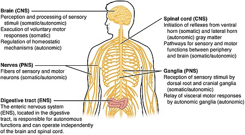
The central nervous system (CNS) serves as the primary integration and coordination center for the body. In bilaterally symmetric and triploblastic animals—which include all multicellular animals except sponges and diploblasts—the CNS integrates received information to influence the activity of all body parts. While basic precursor structures exist in organisms like lancelets and gastropods, true brains are found only in vertebrates, cephalopods, and arthropods.
In vertebrates, the CNS is composed primarily of the brain and the spinal cord. Interestingly, the retina, optic nerve, olfactory nerves, and olfactory epithelium are also technically part of the CNS because they connect directly to brain neurons without intermediate ganglia.
Key Facts
- Primary Components: Consists of the brain and spinal cord in vertebrates.
- Protection: Enclosed by the meninges and bathed in cerebral spinal fluid.
- Composition: Made of nervous tissue and supporting non-nervous cells called neuroglia (glia).
- Unique Access: The olfactory epithelium is the only CNS tissue in direct contact with the environment, bypassing the meninges barrier.
- Mammalian Feature: Only mammals possess a neocortex, the outermost part of the cerebral cortex used for higher thinking.
Anatomical Protection and Environment
The vertebrate CNS is housed within the dorsal body cavity. The brain is protected by the skull in the cranial cavity, while the spinal cord resides within the spinal canal of the vertebrae. Both are wrapped in the meninges, which act as a chemical barrier protecting the brain from neurotoxins found in food.
Rather than standard body fluid, the brain and spinal cord are bathed in cerebral spinal fluid. The spaces between neurons are filled with neuroglia (or glia), which provide essential structural and metabolic support.
Structural Organization
White and Gray Matter
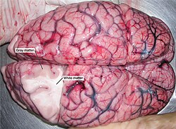
The CNS is characterized by two distinct types of tissue: gray matter and white matter. Gray matter consists of neuron bodies and is found in the cortex and various subcortical nuclei. White matter consists of tracts and commissures that allow for communication between different regions of the system.
The Spinal Cord
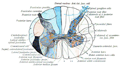
The spinal cord extends from the base of the skull (via the foramen magnum) to approximately the first or second lumbar vertebra. It serves as a conduit for 31 pairs of spinal nerves that connect the CNS to the skin, joints, and muscles.
- Afferent signals: Sensory information traveling from the periphery to the CNS.
- Efferent signals: Motor commands traveling from the CNS to the muscles.
While the spinal cord can process certain reflexes and locomotion, it primarily relays information through spinal tracts to the thalamus and eventually the cerebral cortex.
Cranial Nerves
In addition to spinal nerves, 12 pairs of cranial nerves emerge from the head and neck region. These nerves manage sensory and motor functions for the face and specific muscles, such as the trapezius. The tenth cranial nerve is particularly vital for the autonomic control of internal organs.
The Brain: The Major Processing Unit
The brain is the largest and most complex part of the CNS. It is divided into several specialized regions:
Brainstem
Comprising the medulla, pons, and midbrain, the brainstem is the gateway for motor and autonomic pathways. The medulla is functionally similar to the spinal cord, while the midbrain contains nuclei for the visual and auditory systems and coordinates automatic eye movements.
Cerebellum
The cerebellum contains more neurons than any other brain structure. It is primarily responsible for processing sensory stimuli, motor information, and balance data from the vestibular organ.
Diencephalon
This region includes the thalamus and hypothalamus. The thalamus acts as a sophisticated sorting station, directing incoming sensory information (except smell) to the cerebral hemispheres and linking the cerebrum with the basal ganglia and cerebellum.
Cerebrum
The cerebrum consists of two cerebral hemispheres containing the cortex, basal ganglia, amygdala, and hippocampus. This area governs cognitive capabilities, memory, emotion, perception, and complex motor functions.
CNS vs. Peripheral Nervous System (PNS)
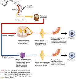
The primary difference between the CNS and PNS lies in their cellular composition and insulation. Both use myelin sheaths to speed up electrical signals, but they use different cells to create them: oligodendrocytes in the CNS and Schwann cells in the PNS. Because peripheral nerves can be over a meter long (e.g., nerves reaching the big toe), they require more extensive myelination than the typically shorter axons found in the CNS.
Development and Evolution
Embryonic Development
The CNS originates from the neural tube. The rostral (front) end differentiates into three vesicles: the prosencephalon, mesencephalon, and rhombencephalon. These further divide into the telencephalon, diencephalon, metencephalon, and myelencephalon, eventually forming the complex structures of the adult brain.
Evolutionary Progression
The CNS has evolved in complexity across different phyla:
- Planarians: Possess a simple CNS consisting of two fused anterior ganglia and longitudinal nerve cords.
- Arthropods: Feature a ventral nerve cord and supra/subesophageal ganglia.
- Mammals: The only vertebrates with a neocortex. The complexity of this region varies; for example, the neocortex of a mouse is roughly 1/100 the size of a monkey's, which is in turn 1/10 the size of a human's. High levels of convolution (gyri and sulci) are found in placental mammals and are particularly extreme in dolphins to support complex echolocation.
| Region | Key Components | Primary Function |
|---|---|---|
| Brainstem | Medulla, Pons, Midbrain | Autonomic control, relay pathways |
| Cerebellum | Neuronal clusters | Balance, motor coordination |
| Diencephalon | Thalamus, Hypothalamus | Information sorting, homeostasis |
| Cerebrum | Cortex, Basal Ganglia, Hippocampus | Cognition, memory, emotion |
| Spinal Cord | White/Gray matter tracts | Signal relay, reflex processing |
Frequently Asked Questions
What is the difference between gray and white matter?
Gray matter consists primarily of neuron cell bodies and is where processing occurs. White matter consists of myelinated axons that form tracts, acting as the communication cables that connect different gray matter regions.
Why is the retina considered part of the CNS?
The retina is technically part of the CNS because it develops from the neural tube and connects directly to the brain's neurons without passing through intermediate ganglia.
What is the role of the neocortex in mammals?
The neocortex is the outermost layer of the cerebral cortex. It is responsible for higher-order thinking and the advanced processing of sensory information.
How does the CNS differ from the PNS in terms of insulation?
While both use myelin for insulation, the CNS uses oligodendrocytes to create these sheaths, whereas the PNS uses Schwann cells. Peripheral nerves generally require more insulation due to their greater length.
What is the function of the meninges?
The meninges are protective membranes that enclose the brain and spinal cord, providing a barrier against chemicals and neurotoxins present in the blood.