Red Blood Cells: The Body's Oxygen Transport System
Red blood cells, also known as erythrocytes, are the most abundant cells in the human body. These specialized cells act as the primary vehicle for transporting oxygen from the lungs to the rest of the body's tissues. In a healthy adult, approximately 84% of all cells are red blood cells, totaling between 20 and 30 trillion units. These cells make up a significant portion of blood volume, accounting for roughly 40% to 45% of the total.
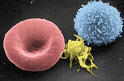
The efficiency of these cells is a result of their unique design. Mature human red blood cells are flexible, biconcave disks that lack a nucleus and other organelles. This absence of internal structures maximizes the space available for hemoglobin, the iron-rich protein that binds to oxygen. Essentially, a red blood cell is a specialized sack of hemoglobin enclosed by a plasma membrane.
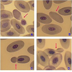
![There is an immense size variation in vertebrate red blood cells, as well as a correlation between cell and nucleus size. Mammalian red blood cells, which do not contain nuclei, are considerably smaller than those of most other vertebrates.[10]](/images/c6/50/c65006049799a6adc9170d493a958598571e680dbde09f1aca6961389de98083.jpg)
Key Facts
- Production Rate: Human adults produce approximately 2.4 million new erythrocytes every second.
- Lifespan: Most cells circulate for 100 to 120 days before being recycled by macrophages.
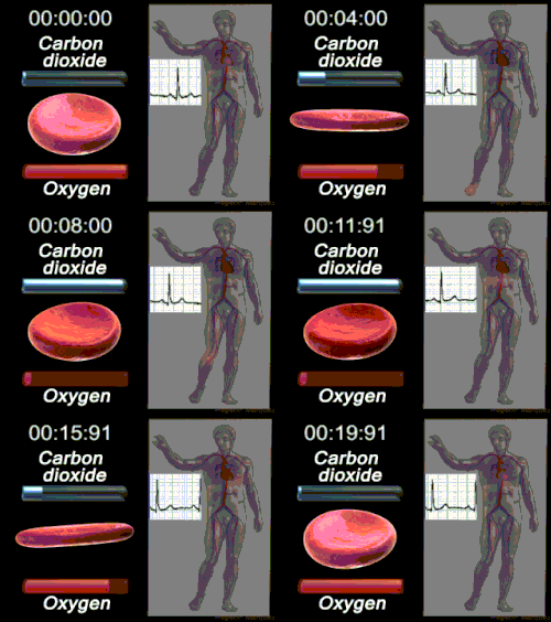
- Circulation Time: A single trip through the circulatory system takes about 60 seconds.
- Cell Count: Men typically have 5–6 million cells per microliter, while women have 4–5 million.
- Physical Size: They average 6.2–8.2 μm in diameter and 2–2.5 μm in maximum thickness.
Structure and Composition
Physical Dimensions
The biconcave shape of the human red blood cell is critical for its function. With a center thickness of only 0.8–1 μm and an average volume of 90 fL, the cell is significantly smaller than most other human cells. This shape provides a high surface-area-to-volume ratio (approximately 136 μm²), which facilitates the rapid exchange of gases. Furthermore, their flexibility allows them to deform and squeeze through narrow capillaries.
Membrane Architecture
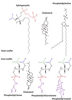
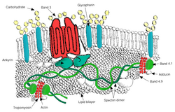
The plasma membrane is a complex bilayer of lipids and proteins that maintains the cell's integrity and regulates transport. The outer monolayer is primarily composed of phosphatidylcholine (PC), while the inner monolayer contains phosphatidylethanolamine (PE), phosphatidylserine (PS), and small amounts of phosphoinositol (PI).
Essential Membrane Proteins
Proteins embedded in the membrane serve various critical roles, categorized primarily by their function:
- Transport Proteins: These include Aquaporin 1 (water), Glut1 (glucose), MCT1 (lactic acid), and various ion exchangers like the Na/K-ATPase and Na-H exchanger.
- Cell Adhesion and Identification: Proteins such as RhD/RhCE (defining the Rh Blood Group), Glycophorin C and D (Gerbich Blood Group), and ICAM-4 help the cell interact with other biological structures.
![Red blood cell membrane proteins separated by SDS-PAGE and silverstained[32]](/images/de/9b/de9ba4efc3f9756a7b7cf226879bd25fde00c43af03af85ac429b8ad562d8e51.jpg)
Biological Functions
Oxygen and Carbon Dioxide Transport
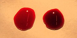
While the primary role of the erythrocyte is oxygen transport, it is equally vital in managing carbon dioxide (CO₂). CO₂ diffuses from tissue cells into the blood, where it is transported back to the lungs. This process is facilitated by carbonic anhydrase, an enzyme that catalyzes the reaction between CO₂ and water. In erythrocytes, CO₂ is transported as bicarbonate (82%), carbamino compounds (11%), and in physical solution (7%).
Secondary Roles
Beyond gas exchange, red blood cells contribute to the immune response and vascular regulation. Research indicates they can aggressively attack bacteria using respiratory protein-generated reactive oxygen species. They also play a role in vasodilation (the widening of blood vessels) through the release of signals like nitric oxide.
The Life Cycle of an Erythrocyte
Erythropoiesis
The process of creating red blood cells is called erythropoiesis. This journey from a committed stem cell in the bone marrow to a mature red blood cell takes approximately seven days. During development, the nucleus is expelled to make room for hemoglobin.
Senescence and Recycling
After 100 to 120 days of service, the cells undergo senescence (biological aging). They lose their flexibility and are eventually identified and removed from circulation by macrophages, which recycle the cell's components.
Clinical Significance and Variations
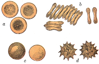
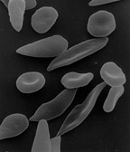
Various conditions can alter the shape and function of red blood cells, a phenomenon generally termed poikilocytosis. For example, in sickle-cell disease, cells take on a crescent shape that can damage internal organs. Additionally, osmotic pressure—the movement of water across the membrane—can cause cells to swell into spheres or shrink and become spiky (crenate).
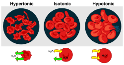
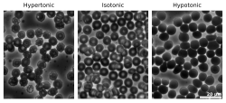
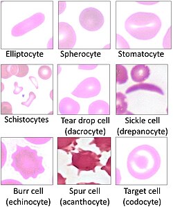
Historically, these cells were first described in 1658 by Jan Swammerdam using a microscope to study frog blood, followed by a more precise description by Anton van Leeuwenhoek in 1674.
| Characteristic | Value/Detail |
|---|---|
| Average Diameter | 6.2–8.2 μm |
| Average Volume | 90 fL |
| Lifespan | 100–120 days |
| Production Rate | ~2.4 million cells/second |
| Primary Protein | Hemoglobin |
| Key Enzyme | Carbonic anhydrase |
Frequently Asked Questions
Why do red blood cells lack a nucleus?
They expel their nucleus during development to create maximum internal space for hemoglobin, allowing the cell to carry the largest possible amount of oxygen.
How does altitude affect red blood cell count?
People living at high altitudes, where oxygen tension is lower, typically produce a higher number of red blood cells to compensate for the decreased oxygen availability.
What is the difference between red blood cells in mammals and other vertebrates?
Mammalian red blood cells are generally smaller and lack nuclei, whereas most other vertebrates have larger red blood cells that retain their nuclei.
What happens to red blood cells at the end of their life?
Once they reach the end of their 100–120 day lifespan, they are removed from the bloodstream and their components are recycled by macrophages.
What is poikilocytosis?
Poikilocytosis is a general term used to describe abnormal variations in the shape of red blood cells, which can be caused by various diseases or osmotic pressure changes.