Ovules: The Biological Foundation of Seed Development
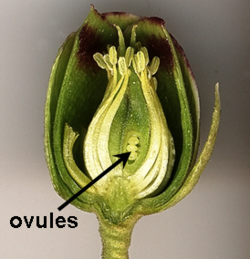
In the complex world of plant reproduction, the ovule serves as the essential structure that houses and gives rise to female reproductive cells. Found within the ovary of flowering plants, these microscopic powerhouses are responsible for the eventual creation of seeds, ensuring the continuation of plant species through generations. Whether in the ancient lineages of gymnosperms or the diverse flowering angiosperms, the ovule is a masterpiece of biological engineering.
Key Facts
- Primary Function: The ovule contains the female gametophyte and develops into a seed after fertilization.
- Core Components: It consists of the integument (outer layer), the nucellus (megasporangium remnant), and the megagametophyte.
- Nutrient Supply: The funicle is the stalk that attaches the ovule to the placenta, providing vital nourishment.
- Fertilization Entry: The micropyle is a small opening in the integuments that allows pollen to enter.
- Angiosperm Uniqueness: Flowering plants undergo double fertilization, creating both a zygote and a nutrient-rich endosperm.
Anatomy and Structure of the Ovule
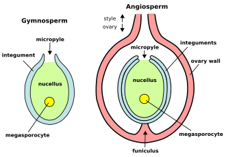
An ovule is composed of several specialized parts that work in harmony to facilitate reproduction. The outermost protective layer is known as the integument. In gymnosperms, there is typically one integument (unitegmic), whereas angiosperms usually possess two (bitegmic). These integuments eventually harden to become the protective seed coat.
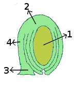
Inside the integuments lies the nucellus, a layer of diploid maternal tissue that functions as the megasporangium. Within the nucellus, a single cell called the megasporocyte undergoes meiosis to produce megaspores. In most cases, only one functional megaspore survives to develop into the megagametophyte (also known as the embryo sac in angiosperms).
Key Anatomical Terms
- Micropyle: An opening at the apex of the integuments that allows the pollen tube to reach the female cells.
- Chalaza: The region opposite the micropyle where the nucellus joins the integuments.
- Funicle: The stalk connecting the ovule to the plant's placenta.
- Raphe: A structural ridge often associated with the funicle.
Placentation: How Ovules are Positioned
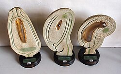
The way ovules are attached to the ovary wall is known as placentation. This arrangement varies significantly across different plant species:
- Apical: The placenta is located at the very top (apex) of the ovary.
- Axile: The ovary is divided into segments, with placentae located along the central axis where carpel margins meet (e.g., Citrus, Hibiscus).
- Basal: The placenta sits at the bottom of the ovary on a protrusion of the receptacle (e.g., Sunflower).
- Free-central: Ovules are attached to a central axis that is not connected to the ovary walls (e.g., Dianthus).
- Marginal: Ovules are attached along a single ridge on one side of the ovary, common in legumes like peas.
- Parietal: Placentae are located on the inner walls of the ovary (e.g., Brassica).
- Superficial: Similar to axile, but the placentae are on the inner surfaces of a multi-chambered ovary.
Development and Fertilization
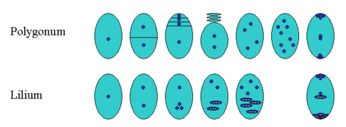
The development of the female gametophyte follows different patterns depending on the species. In the common Polygonum type, a single functional megaspore undergoes three rounds of mitosis to form the embryo sac. In contrast, some species like Lilium follow a Fritillaria type, where multiple nuclei fuse to create different ploidy levels.
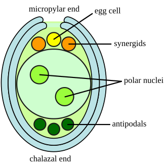
In angiosperms, the embryo sac typically consists of seven cells and eight nuclei. These include the egg cell, two synergids (which guide the pollen tube), three antipodal cells, and a large central cell containing two polar nuclei.
Fertilization in flowering plants is a unique process called double fertilization. One sperm nucleus fuses with the egg to form a diploid zygote, which becomes the embryo. A second sperm nucleus fuses with the two polar nuclei in the central cell to create the endosperm. The endosperm acts as a food reservoir—similar to a yolk—providing starch, proteins, and oils to the developing seedling.
Summary of Ovule Components and Functions
| Structure | Description | Primary Function |
|---|---|---|
| Integument | Outer protective layers | Forms the seed coat |
| Nucellus | Internal diploid tissue | Houses the megasporocyte |
| Micropyle | Small opening at the apex | Entry point for pollen tube |
| Megagametophyte | Haploid female gametophyte | Produces the egg cell |
| Endosperm | Triploid tissue (in angiosperms) | Nutrient storage for embryo |
Frequently Asked Questions
What is the difference between gymnosperm and angiosperm ovules?
Gymnosperm ovules are typically borne on the surface of scales within cones and produce archegonia to house egg cells. Angiosperm ovules are enclosed within an ovary and undergo double fertilization to produce both an embryo and endosperm.
What is the function of the micropyle?
The micropyle is a specialized opening that allows the pollen tube to enter the ovule to reach the egg cell. During germination, it also serves as the exit point for the seedling's radicle.
How does the endosperm help the plant?
The endosperm serves as a vital food source, containing essential nutrients like starch, oils, and proteins that nourish the developing embryo and the young seedling until it can perform photosynthesis.
What is double fertilization?
Double fertilization is a process unique to angiosperms where one sperm nucleus fuses with the egg to form a zygote, while a second sperm nucleus fuses with the polar nuclei to form the endosperm.
What is the role of the funicle?
The funicle is a stalk-like structure that attaches the ovule to the placenta within the ovary, facilitating the transfer of nutrients to the developing ovule.