Notochord: The Evolutionary Blueprint of Chordate Development
The notochord is a defining biological feature of all chordates. This elastic, rod-like structure serves as a primary axial support and a critical signaling center during early development. While it remains a permanent structural element in some primitive chordates, in vertebrates, it acts as a temporary scaffold that guides the formation of the complex vertebral column.
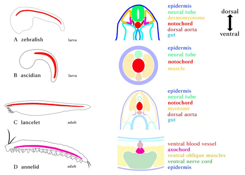
Derived from the axial mesoderm (the middle layer of embryonic cells), the notochord is positioned along the body's head-to-tail axis. It sits dorsal to the gut tube and ventral to the dorsal nerve cord, providing a midline reference for the surrounding tissues as the embryo grows.
Key Facts
- Defining Feature: The notochord is the synapomorphy (shared derived characteristic) that defines the phylum Chordata.
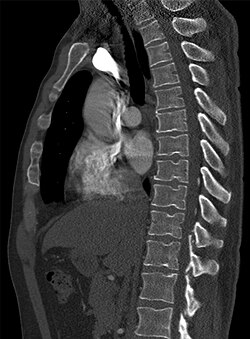
- Vertebrate Fate: In most vertebrates, it is replaced by the vertebral column, persisting only as the nucleus pulposus in intervertebral discs.
- Chemical Composition: It consists of a glycoprotein core encased in two helical collagen-elastin sheaths.
- Signaling Role: It secretes Sonic Hedgehog (SHH), a protein essential for patterning the central nervous system.
- Locomotion: In aquatic animals, it prevents the body from telescoping, enabling side-to-side swimming motions.
Structure and Mechanical Function
The notochord is engineered for both flexibility and strength. Its core is composed of vacuolated, turgid cells filled with glycoproteins. These cells are wrapped in a sheath of collagen fibers wound into two opposing helices. The specific angle of these fibers determines the mechanical response of the rod: depending on the internal pressure, the notochord will either shorten and thicken or lengthen and thin.
This structural rigidity is vital for locomotion in early chordates. By providing a stiff axis, the notochord allows muscle fibers attached to its sides to contract alternately. This creates a sculling motion that propels the animal through water, preventing the body from collapsing like an earthworm during movement.
The Process of Notogenesis and Signaling
Notogenesis is the developmental process by which the notochord forms. It begins during gastrulation, where cells migrate from the primitive node and pit to form a solid rod of polygonal cells. This process is closely synchronized with neurulation, the formation of the neural tube.
Beyond its physical support, the notochord acts as a chemical command center. It secretes a morphogen called Sonic Hedgehog (SHH). This protein establishes the ventral pole of the embryo's dorsal-ventral axis. In neurology, SHH is critical for the development of motor neurons; without the signaling from the notochord, the ventral neural tube would not develop the necessary motor functions.
Presence Across Different Species
The persistence of the notochord varies significantly across the chordate lineage:
- Cephalochordates (Lancelets): The notochord persists throughout adulthood as the primary body support. In lancelets, it even extends beyond the neural tube to assist in burrowing into sediment.
- Tunicates: The notochord is present only during the larval stage and is lost as the animal reaches adulthood.
- Vertebrates: Except for the hagfish, the notochord is an embryonic structure. It is eventually replaced by the bony or cartilaginous vertebral column.
Post-Embryonic Retention
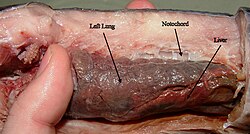
While most vertebrates transition to a spine, several groups retain a post-embryonic notochord, including lampreys, hagfish, coelacanths, lungfish, and sturgeons.
| Group | Persistence | Adult Role/Fate |
|---|---|---|
| Lancelets | Lifelong | Main structural support and burrowing aid |
| Tunicates | Larval only | Absent in adult stage |
| Most Vertebrates | Embryonic | Becomes nucleus pulposus of intervertebral discs |
| Hagfish/Lamprey | Lifelong | Primary axial support |
Clinical Significance in Humans
In humans, the notochord is almost entirely replaced by chondrocyte-like cells by age four. However, remnants remain in the nucleus pulposus of the spinal discs. If these cells do not regress or migrate correctly, several conditions can arise:
- Persistent Notochordal Canal: A condition where notochordal cells persist within the vertebra.
- Tornwaldt Cyst: A depression or cyst forming in the nasopharynx if the notochord fails to separate properly.
- Chordoma: A rare type of cancer originating from notochordal remnants.
Evolutionary Origins
The origin of the notochord is a subject of ongoing scientific debate. Two primary theories exist: first, that it evolved de novo in chordates through new combinations of developmental programs; and second, that it derived from a homologous muscular structure called the axochord found in annelid-like ancestors. Evidence from gene expression patterns supports the axochord theory, though the possibility of convergent evolution (homoplasy) remains.
Frequently Asked Questions
What happens to the notochord in adult humans?
In humans, the notochord mostly disappears during development, with its remnants forming the nucleus pulposus, the gel-like center of the intervertebral discs.
How does the notochord affect the nervous system?
The notochord secretes the protein Sonic Hedgehog (SHH), which signals the developing neural tube to form motor neurons on its ventral side.
Which animals keep their notochord for their entire lives?
Lancelets (Amphioxus), hagfish, lampreys, and some fish like sturgeons and lungfish retain the notochord into adulthood.
What is the difference between a notochord and a vertebral column?
The notochord is a flexible, rod-like structure made of glycoproteins and collagen, whereas the vertebral column is a segmented series of bony or cartilaginous vertebrae that provide more rigid support.
What is a chordoma?
A chordoma is a rare malignancy that arises from the remnants of the embryonic notochord that failed to regress.