Heart Valves: Anatomy, Function, and Clinical Significance
The heart is a complex muscular pump designed to keep blood moving in a single, efficient direction throughout the body. Central to this process are the heart valves—biological one-way gates that ensure blood flows forward and prevent dangerous backflow. In mammals, the heart typically utilizes four primary valves that open and close in response to pressure differences between the heart's chambers and the connecting arteries.
These valves are lined with endocardium (the inner lining of the heart) and are situated around the fibrous rings of the cardiac skeleton. They operate using flaps called leaflets or cusps, which act similarly to a duckbill valve: they are pushed open by blood pressure to allow flow and snap shut to seal the passage.

Key Facts
- Primary Function: Ensure unidirectional blood flow through the heart chambers.
- Mechanism: Operated by pressure differentials on either side of the valve.
- Valve Types: Divided into two atrioventricular (AV) valves and two semilunar (SL) valves.
- Heart Sounds: The "lub-dub" sound is created by the closing of these valves.
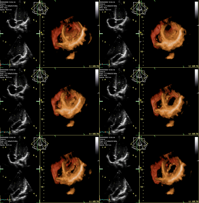
- Diagnosis: Echocardiography (ultrasound) is the primary tool for diagnosing valve dysfunction.
The Four Primary Heart Valves
The valves are categorized based on their location and the specific chambers they separate.
Atrioventricular (AV) Valves
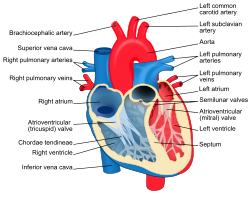
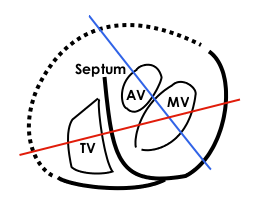
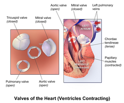
Located between the upper atria and the lower ventricles, these valves prevent blood from flowing backward into the atria during systole (the contraction phase of the heart). They are uniquely anchored to the ventricle walls by chordae tendineae, which act like tethering cords to prevent the valves from inverting under high pressure.
- Mitral Valve (Bicuspid Valve): Located on the left side of the heart. It has two cusps and allows blood to flow from the left atrium into the left ventricle.
- Tricuspid Valve: Located on the right side of the heart. It has three cusps and separates the right atrium from the right ventricle.
Semilunar (SL) Valves
These valves are located at the exits of the ventricles, leading into the major arteries. Unlike AV valves, they do not have chordae tendineae and are shaped like half-moons.
- Aortic Valve: Located between the left ventricle and the aorta; it allows oxygenated blood to exit the heart to the rest of the body.
- Pulmonary Valve: Located between the right ventricle and the pulmonary artery; it directs blood toward the lungs.
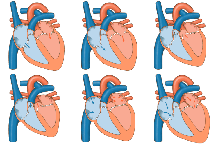
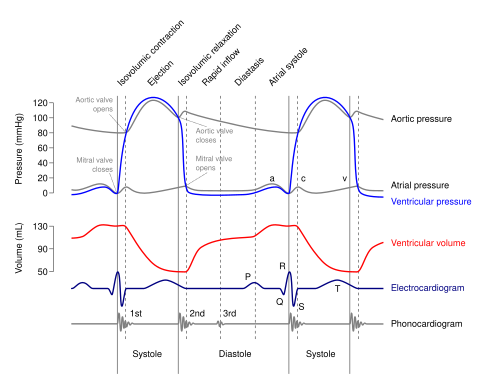
The coordinated movement of these valves is essential for the cardiac cycle. The closure of the AV valves produces the first heart sound (S1, "lub"), while the closure of the SL valves produces the second heart sound (S2, "dub").
Summary of Heart Valve Characteristics
| Valve Name | Type | Number of Cusps | Location | Prevents Backflow Into... |
|---|---|---|---|---|
| Tricuspid | Atrioventricular | 3 | Right Atrium & Ventricle | Right Atrium |
| Mitral (Bicuspid) | Atrioventricular | 2 | Left Atrium & Ventricle | Left Atrium |
| Pulmonary | Semilunar | 3 | Right Ventricle & Pulmonary Trunk | Right Ventricle |
| Aortic | Semilunar | 3 | Left Ventricle & Aorta | Left Ventricle |
Physiology and Development
The motion of heart valves is governed by fluid dynamics, often analyzed using the Navier–Stokes equation, which considers blood pressure, pericardial fluid, and external loading. During diastole (the relaxation phase), the mitral valve opens as atrial pressure exceeds ventricular pressure, allowing blood to fill the ventricle—a process known as preloading.
Developmentally, the AV valves form from the invagination of the atrioventricular canals and the extension of the septum intermedium. The semilunar valves originate from endocardial cushions—four thickenings at the cardiac end of the truncus arteriosus. By the ninth week of embryonic development, these valves are visible as unique structures.
Clinical Significance and Valvular Disease
When valves fail to function correctly, it results in valvular heart disease. This generally manifests in two forms:
- Regurgitation (Insufficiency): The valve does not close tightly, allowing blood to leak backward.
- Stenosis: The valve becomes thickened or narrow, restricting blood flow.
Common conditions include mitral valve prolapse, where connective tissue weakens and the valve cusp displaces into the left atrium during systole. Other causes of dysfunction include infective endocarditis (bacterial infection of the valve lining) and rheumatic fever, which often leads to mitral stenosis.
Congenital Defects
Some valve issues are present from birth. The most common is the bicuspid aortic valve, where two cusps fuse during development. This often leads to calcific aortic stenosis much earlier in life than in those with a tricuspid valve. Other rare defects include tricuspid atresia (absence of the valve) and Ebstein's anomaly (displacement of the tricuspid septal leaflet).
Frequently Asked Questions
What is the difference between stenosis and regurgitation?
Stenosis occurs when a valve becomes narrow or stiff, making it difficult for blood to flow forward. Regurgitation occurs when a valve fails to close completely, allowing blood to leak backward into the previous chamber.
What causes the "lub-dub" sound of the heart?
The "lub" (S1) is the sound of the mitral and tricuspid valves closing at the start of ventricular contraction. The "dub" (S2) is the sound of the aortic and pulmonary valves closing as the ventricles relax.
How are heart valve problems diagnosed?
The primary diagnostic tool is echocardiography, which uses ultrasound waves to create images of the heart's structure and blood flow in real-time.
Can damaged heart valves be fixed?
Yes, depending on the severity, damaged valves can be surgically repaired or replaced with artificial heart valves, such as the historically significant Starr-Edwards valve or modern pericardial valves.
What is a bicuspid aortic valve?
It is a congenital heart defect where the aortic valve has only two leaflets instead of the usual three, which can increase the risk of developing aortic stenosis earlier in life.