Heart Atria: Anatomy, Function, and Clinical Significance
The atrium (plural: atria) serves as the essential "entry hall" of the heart. These two upper chambers are the first point of contact for blood returning to the heart from the rest of the body and the lungs. By acting as receiving reservoirs, the atria ensure that blood flows smoothly and continuously into the ventricles, the heart's primary pumping chambers.
In the human heart, the atria operate in coordination with the ventricles during the cardiac cycle. They receive blood while relaxed during diastole and contract during systole to push blood through the atrioventricular valves. While often overshadowed by the powerful ventricles, the atria are critical for maintaining efficient cardiac output and regulating blood volume.
Key Facts
- Two Chambers: The heart contains a right atrium (receives deoxygenated blood) and a left atrium (receives oxygenated blood).
- Blood Flow: Blood moves from the atria to the ventricles via the tricuspid valve (right) and the mitral valve (left).
- Cardiac Output: Atria allow for approximately 75% more cardiac output by preventing the interruption of venous flow.
- Pacemaker Location: The sinoatrial (SA) node, which triggers the heartbeat, is located in the right atrium.
- Clinical Risk: The left atrial appendage is a common site for blood clot formation during atrial fibrillation.
Anatomy of the Atria
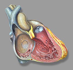
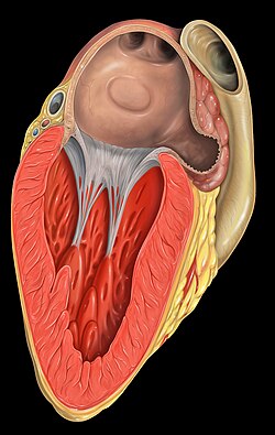
The human heart is a four-chambered organ. The right atrium and right ventricle together form the right heart, while the left atrium and left ventricle form the left heart. Unlike the ventricles, the atria do not have valves at their inlets, which allows venous pulsations to be detected in the jugular vein (known as jugular venous pressure).
Internally, the atria feature pectinate muscles (rough muscular ridges) and the crista terminalis, a boundary within the chamber. The right atrium also contains the sinus venarum, a smooth-walled area derived from the embryonic sinus venosus that surrounds the openings of the venae cavae and the coronary sinus.
The Right Atrium
The right atrium is responsible for collecting deoxygenated blood from the superior vena cava, inferior vena cava, coronary sinus, and various cardiac veins. It then pumps this blood through the tricuspid valve into the right ventricle, which subsequently sends it to the lungs via the pulmonary artery.
A notable feature is the right atrial appendage (or right auricle), a wedge-shaped pouch on the front upper surface. The right atrium is separated from the left by the interatrial septum, which features a depression called the fossa ovalis.
The Left Atrium
The left atrium receives oxygenated blood from the left and right pulmonary veins. This blood is pumped through the mitral valve (the left atrioventricular valve) into the left ventricle, which then distributes it to the rest of the body through the aorta.
The left atrium is primarily supplied with blood by the left circumflex coronary artery. Its venous drainage is partly handled by the oblique vein of the left atrium.
The Left Atrial Appendage (LAA)
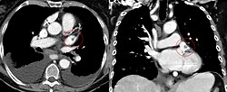
The left atrial appendage is a muscular, ear-shaped pouch located in the upper part of the left atrium. It is characterized by a tubular, trabeculated structure and is thought to act as a decompression chamber when left atrial pressure is high. Additionally, it helps modulate blood volume by secreting natriuretic peptides, such as atrial natriuretic peptide (ANP) and brain natriuretic peptide (BNP).
Medical imaging, such as CT scans, categorizes LAA morphology into four groups: chicken wing (48%), cactus (30%), windsock (19%), and cauliflower (3%). The cauliflower shape is most frequently associated with embolism.
In conditions like atrial fibrillation—where the atrium quivers instead of contracting—blood can stagnate in the LAA, leading to the formation of thrombi (blood clots). If these clots dislodge, they can cause a stroke. Consequently, surgeons may perform a left atrial appendage occlusion to prevent this risk.
Physiological Function and Conduction
The primary role of the atria is to facilitate uninterrupted venous flow. Because they are distensible and lack inlet valves, they prevent the "back-up" of blood that would occur during ventricular systole. This prevents circulatory inertia and significantly increases overall cardiac output.
The atria also contain low-pressure baroreceptors (volume receptors). When these detect a drop in atrial pressure, they signal the hypothalamus to release vasopressin to help maintain blood volume.
The Electrical System
The heart's rhythm begins in the sinoatrial (SA) node, located in the posterior right atrium. These pacemaker cells spontaneously depolarize, creating an action potential that spreads across both atria, causing them to contract. The signal then reaches the atrioventricular (AV) node, located between the atria and ventricles, to coordinate the subsequent ventricular contraction.
Development and Clinical Disorders
During embryonic development, the atrium begins as a single chamber. Around the fourth week, the septum primum divides it into left and right chambers. In the fetus, an opening called the foramen ovale allows blood to bypass the lungs. This normally closes at birth, leaving the fossa ovalis.
If this opening fails to close, it is called a patent foramen ovale (PFO), an atrial septal defect present in about 25% of the population. While often harmless, it can occasionally lead to paradoxical embolization and stroke.
| Feature | Right Atrium | Left Atrium |
|---|---|---|
| Blood Type | Deoxygenated | Oxygenated |
| Primary Source | Venae cavae & Coronary sinus | Pulmonary veins |
| Exit Valve | Tricuspid Valve | Mitral Valve |
| Destination | Right Ventricle | Left Ventricle |
| Key Feature | SA Node / Fossa ovalis | Left Atrial Appendage (LAA) |
Frequently Asked Questions
What is the difference between an atrium and an auricle?
In modern human anatomy, the atrium is the entire upper chamber of the heart, while the auricle (or atrial appendage) is the small, ear-shaped muscular pouch attached to the atrium. In some other animals, such as mollusks, the term auricle is still used to describe the chamber itself.
Why is the left atrial appendage dangerous during atrial fibrillation?
During atrial fibrillation, the LAA does not contract effectively, causing blood to pool and stagnate. This stasis predisposes the heart to the formation of blood clots (thrombi), which can travel to the brain and cause an ischemic stroke.
What happens if a person has an atrial septal defect?
An atrial septal defect allows blood to flow in the reverse direction—from the left atrium to the right. This can reduce overall cardiac output and, if severe or untreated, may lead to heart failure or cardiac arrest.
How do the atria increase cardiac output?
By acting as reservoirs that accept continuous venous flow even while the ventricles are contracting, the atria prevent circulatory inertia. This mechanism allows for approximately 75% more cardiac output than would be possible if veins ended directly at the ventricular valves.
What is the role of the SA node in the atria?
The sinoatrial (SA) node acts as the heart's natural pacemaker. Located in the right atrium, it generates the electrical impulses that trigger the atria to contract, ensuring blood is efficiently moved into the ventricles before they pump.