Aorta: Anatomy, Function, and Clinical Significance
The aorta is the largest and most critical artery in the human body. Originating from the left ventricle of the heart, it serves as the primary trunk of the systemic circulation, distributing oxygen-rich blood to nearly every tissue and organ in the body, with the exception of the respiratory zone of the lungs.
From its start at the heart, the aorta travels upward, curves sharply, and then descends through the chest and abdomen, eventually splitting into smaller arteries to supply the lower extremities. Its unique elastic structure allows it to manage the high-pressure output of the heart, ensuring a steady flow of blood throughout the body.
Key Facts
- Primary Role: Distributes oxygenated blood to the entire systemic circulation.
- Origin: Begins at the aortic valve of the left ventricle.
- Termination: Ends at the aortic bifurcation, splitting into the common iliac arteries.
- Structure: An elastic artery composed of three layers: tunica intima, tunica media, and tunica externa.
- Key Mechanism: Utilizes the Windkessel effect to maintain blood pressure during the heart's relaxation phase (diastole).
Anatomical Structure and Sections
To simplify its complex path, anatomists typically divide the aorta into sections based on either the anatomical compartment it occupies or the direction of blood flow.
Compartmental Division
- Thoracic Aorta: The portion running from the heart to the diaphragm.
- Abdominal Aorta: The portion extending from the diaphragm to the aortic bifurcation.
Flow-Based Division
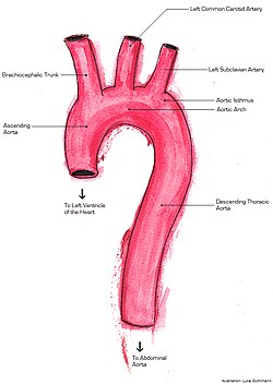
Following the path of blood flow, the aorta is categorized into the ascending aorta, the aortic arch, and the descending aorta (which is further split into thoracic and abdominal parts).
Detailed Regional Anatomy
The Ascending Aorta
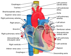
The ascending aorta begins at the aortic valve. It shares a pericardial sheath with the pulmonary trunk, twisting around it to move from a posterior position to the right and anterior side. At its root are the sinuses of Valsalva (aortic sinuses). The left and right coronary sinuses give rise to the coronary arteries, which supply the heart muscle itself, while the posterior sinus is non-coronary.
The Aortic Arch
The aortic arch loops over the left pulmonary artery and the left main bronchus. It is connected to the pulmonary trunk by the ligamentum arteriosum, a remnant of fetal circulation. This section is critical for supplying the upper body via three major branches: the brachiocephalic trunk, the left common carotid artery, and the left subclavian artery.
The Descending Aorta
The descending aorta begins at the level of the fourth and fifth thoracic vertebrae. It is divided into two primary regions:
- Thoracic Part: Supplies the chest wall via intercostal and subcostal arteries, as well as the esophagus and bronchial arteries.
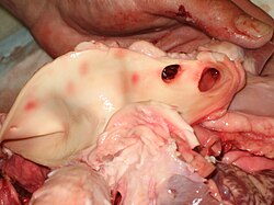
- Abdominal Part: Begins at the aortic hiatus of the diaphragm. It provides blood to vital organs via the celiac trunk, superior and inferior mesenteric arteries, and renal arteries. It terminates by dividing into the common iliac arteries and the median sacral artery.
Microanatomy and Physiology
The aorta is an elastic artery, meaning it is highly distensible. Its wall consists of three layers: the tunica intima (innermost), tunica media (middle), and tunica externa (outermost). Because the wall is so thick, the outer layers are nourished by a network of tiny vessels called vasa vasorum.
The aortic arch also houses baroreceptors and chemoreceptors. These specialized sensors monitor blood pressure, pH, and carbon dioxide levels, sending data to the medulla oblongata in the brain to maintain homeostasis via the autonomic nervous system.
The Windkessel Effect
When the left ventricle contracts, the aorta expands to accommodate the surge of blood. During diastole (the heart's resting phase), the elastic walls recoil. This Windkessel effect converts the pulsatile output of the heart into a more continuous, smooth flow, conserving energy and protecting smaller downstream vessels from pressure spikes.
Clinical Significance and Variations
Due to its size and pressure, the aorta is susceptible to several medical conditions:
- Aneurysms: Abnormal bulging of the aortic wall, which can be caused by genetics, infection (syphilis), or age.
- Dissection: A tear in the inner layer of the aorta that allows blood to flow between the layers of the wall.
- Coarctation: A congenital narrowing of the aorta.
- Atherosclerosis: The buildup of fats and cholesterol on the artery walls.
Anatomical variations also occur. In dextrocardia or situs inversus, the aorta may be located on the right side of the body. Additionally, a patent ductus arteriosus occurs when the fetal connection between the pulmonary artery and aorta fails to close after birth.
Summary of Aortic Branches
| Aortic Section | Primary Branches | Regions Supplied |
|---|---|---|
| Ascending Aorta | Right and Left Coronary Arteries | Heart muscle |
| Aortic Arch | Brachiocephalic trunk, L. Common Carotid, L. Subclavian | Head, neck, and arms |
| Thoracic Aorta | Intercostal, Subcostal, Bronchial, Esophageal arteries | Chest wall and thoracic organs |
| Abdominal Aorta | Celiac trunk, Mesenteric, Renal, Gonadal arteries | Abdominal organs and glands |
| Terminal Branches | Common Iliac Arteries, Median Sacral Artery | Pelvis and legs |
Frequently Asked Questions
What is the main function of the aorta?
The aorta's primary function is to carry oxygenated blood from the left ventricle of the heart to the rest of the body's systemic circulation, ensuring all organs and tissues receive necessary oxygen and nutrients.
What is the Windkessel effect?
The Windkessel effect refers to the elastic recoil of the aorta. It expands during heart contraction (systole) and contracts during relaxation (diastole), which smooths out blood flow and maintains a steady blood pressure.
What happens during an aortic dissection?
An aortic dissection occurs when a tear develops in the inner layer (tunica intima) of the aorta, allowing blood to force its way between the layers of the arterial wall, creating a false lumen.
How does the aorta differ in fish compared to humans?
Unlike humans, fish have two separate aortas: a ventral aorta that carries de-oxygenated blood from the heart to the gills, and a dorsal aorta that carries oxygenated blood from the gills to the body.
What are the vasa vasorum?
The vasa vasorum are a network of small blood vessels that supply the outer layers (tunica externa and tunica media) of the aorta, as the vessel wall is too thick for oxygen to diffuse from the main lumen.