Fovea Centralis: The Anatomy of High-Acuity Vision
The fovea centralis is a specialized, small pit located at the center of the macula lutea in the retina. While it occupies a tiny fraction of the eye's total surface area, it is the primary engine for sharp central vision. This high-resolution capability is essential for detailed tasks such as reading, driving, and recognizing faces.
The term "fovea" is derived from the Latin word for "pit," a description that reflects its physical structure as a depression in the inner retinal surface. Named by German histologist Carl Bergmann, the fovea is engineered specifically for maximum visual acuity.
Key Facts
- Primary Function: Responsible for sharp central vision and the ability to distinguish fine detail and color.
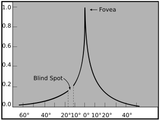
- Cell Composition: Composed almost entirely of densely packed cone photoreceptors, with virtually no rods in the center.
- Avascular Zone: Contains a 0.5 mm diameter area without blood vessels to prevent light dispersion.
- Neural Impact: Despite its small size, approximately 50% of the optic nerve fibers carry information from the fovea.
- Brain Processing: Occupies less than 1% of the retina but utilizes over 50% of the brain's visual cortex.
Anatomical Structure and Regions
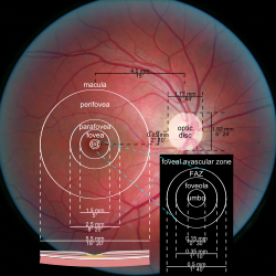
The fovea is not a uniform structure but is organized into concentric regions that vary in cell density and function. The central-most part is the foveola, a pit approximately 0.35 mm in diameter. In this region, cones are packed in a tight hexagonal pattern and are thinner and more rod-like than cones found elsewhere in the eye.
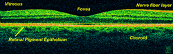
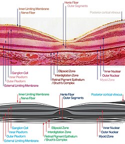
Surrounding the foveola is the foveal rim, which is the thickest part of the retina because it contains neurons displaced from the central pit. To ensure that light reaches the photoreceptors without interference, the fovea features a foveal avascular zone (FAZ). This lack of blood vessels prevents light scattering, meaning the fovea receives most of its oxygen from the choroid, located across the Bruch's membrane and retinal pigment epithelium.
The Macular Hierarchy
The fovea is the core of a larger system known as the macula lutea. This system is divided into three primary zones:
- Parafovea: An intermediate belt extending to a 1.25 mm radius. It contains the highest overall density of cones and a ganglion cell layer (GCL) consisting of more than five layers of cells.
- Perifovea: The outermost region extending to a 2.75 mm radius. Here, visual acuity drops, and the GCL contains only two to four layers of cells. Cone density decreases significantly to 12 per 100 micrometers, compared to 50 per 100 micrometers in the central fovea.
- Peripheral Retina: The area beyond the perifovea, which provides low-resolution, highly compressed visual information.
Visual Function and Properties
The fovea's exceptional acuity is due to a low ratio of ganglion cells to photoreceptors (approximately 2.5). In this region, almost every ganglion cell receives data from a single cone, ensuring that the signal is not "averaged" or blurred, which allows for 20/20 vision.
Because the fovea lacks rods—the photoreceptors responsible for low-light vision—it is not sensitive to dim lighting. This is why astronomers use averted vision (looking slightly to the side of an object) to see dim stars, shifting the image to the rod-rich peripheral retina.
Protection and Color Sensitivity
The fovea contains high concentrations of yellow carotenoid pigments, specifically lutein and zeaxanthin. These pigments are concentrated in the Henle fiber layer (radial axons of photoreceptors) and the cones. They serve two main purposes:
- Protection: They shield sensitive cones from high-intensity blue light damage.
- Acuity Enhancement: They reduce sensitivity to short wavelengths, which counteracts chromatic aberration.
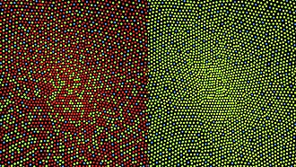
Interestingly, blue-sensitive cones are sparse in the very center of the fovea, with their maximum density occurring in a ring around the center. Consequently, maximum acuity for blue light is lower and occurs about 1° off-center.
Comparative Anatomy and Technical Metrics
In humans, the average focal length of the eye is 17.1 mm. With an average of 147,000 cone cells per square millimeter, a single cone's angle of view is approximately 31.46 arc seconds. This biological "pixel density" informs how we perceive digital screens; for a screen to match average foveal density at a distance of 10 inches, it would require a density of 655.6 PPI.
While humans have a single fovea, some birds—such as hawks, hummingbirds, and kingfishers—possess a double fovea. This includes a temporal fovea that allows them to track slow movements more effectively. Some birds, like the Common Buzzard, can reach cone densities of 1,000,000 per square millimeter.
![Distribution of rods and cones along a line passing through the fovea and the blind spot of a human eye[23]](/images/55/61/5561fa117e31394db3738defafd6013de2de3c439cf3b9d3f5a4e3f372949bc8.webp)
| Region | Diameter/Radius | Key Characteristic | Cone Density |
|---|---|---|---|
| Macula Lutea | 5.5 mm (Dia) | Elliptical shape; GCL > 1 layer | Variable |
| Parafovea | 1.25 mm (Rad) | Highest cone density; GCL > 5 layers | Highest |
| Perifovea | 2.75 mm (Rad) | GCL 2–4 layers; lower acuity | 12 / 100 μm |
| Fovea Centralis | 1.5 mm (Dia) | Central depression; 20/20 vision | Variable |
| Foveola | 0.35 mm (Dia) | Only cones present; no rods | 50 / 100 μm |
| FAZ | 0.5 mm (Dia) | Avascular (no blood vessels) | N/A |
Frequently Asked Questions
What is the difference between the fovea and the optic disc?
The fovea is the center of the macula responsible for sharp central vision, while the optic disc is the area where the optic nerve exits the eye; the optic disc is often referred to as the blind spot because it lacks photoreceptors.
Why can't we see well in dim light using our central vision?
The fovea consists almost entirely of cones, which require significant light to function. Rods, which are sensitive to dim light, are absent from the center of the fovea and are located in the peripheral retina.
What is the foveal avascular zone (FAZ)?
The FAZ is a small region (0.5 mm in diameter) in the center of the fovea that is completely devoid of blood vessels. This prevents blood vessels from scattering or absorbing light, allowing for maximum visual clarity.
How does the fovea contribute to our brain's processing of images?
Although the fovea represents less than 1% of the retina's total area, it is so critical for detail that it occupies over 50% of the visual cortex in the brain.
What role do lutein and zeaxanthin play in the fovea?
These yellow carotenoid pigments protect the fovea from damage caused by high-intensity blue light and improve visual acuity by reducing chromatic aberration.