Esophagus: Anatomy, Function, and Clinical Significance
The esophagus is a vital component of the digestive system, serving as the muscular conduit that transports food and liquids from the pharynx to the stomach. Originating from the embryonic foregut, this tubular structure is more than a simple pipe; it is a complex organ with specific histological layers and regulatory sphincters that ensure the one-way movement of nutrients while protecting the lungs and the esophageal lining from gastric acid.
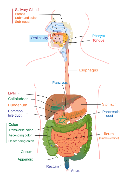
Key Facts
- Primary Function: Transports food from the laryngopharynx to the stomach.
- Key Junction: The gastroesophageal junction (z-line) marks the transition from stratified squamous epithelium to simple columnar epithelium.
- Critical Constrictions: The esophagus has four primary points of narrowing that can be clinically significant.
- Gene Expression: Nearly 70% of the human genome's protein-coding genes are expressed in the normal esophagus.
- Common Malformations: Esophageal atresia and tracheoesophageal fistulas occur in approximately 1 in 3,500 births.
Anatomical Structure and Position
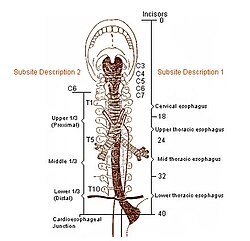
The esophagus is positioned posterior to the trachea and the heart. Its path is defined by several physiological constrictions where the tube is naturally narrowed. These occur at:
- The junction where the laryngopharynx meets the esophagus, located behind the cricoid cartilage.
- The point where the aortic arch crosses the front of the tube in the superior mediastinum.
- The area where the left main bronchus compresses the esophagus in the posterior mediastinum.
- The esophageal hiatus, where the tube passes through the diaphragm.
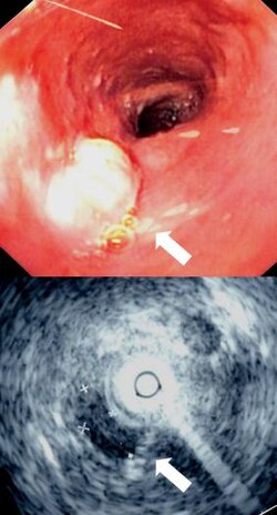
The Gastroesophageal Junction
The lower end of the esophagus meets the stomach at the gastroesophageal junction. This area is characterized by the z-line, an irregular zig-zag line where the pink esophageal mucosa transitions into the deeper red gastric mucosa. Histologically, this represents a shift from stratified squamous epithelium (designed for protection against abrasion) to simple columnar epithelium (designed for secretion and absorption). The functional lower esophageal sphincter is typically located about 3 cm below this z-line.
Microanatomy and Molecular Biology
The esophageal wall consists of four distinct layers. To maintain its integrity and function, the organ expresses a vast array of proteins. Specifically, proteins involved in squamous differentiation, such as keratins KRT13, KRT4, and KRT6C, are highly specific to the esophagus. Additionally, mucins like MUC21 and MUC22 provide essential lubrication for the inner surface to facilitate the smooth passage of food.
Clinical Significance and Pathology
Various conditions can impair the function of the esophagus, ranging from inflammation to structural malformations.
Inflammation (Esophagitis)
Esophagitis is the inflammation of the esophageal lining. Common causes include the reflux of gastric acids, infections (such as Candida albicans in immunocompromised individuals), ingested corrosives, certain medications like bisphosphonates, and food allergies. Eosinophilic esophagitis is a specific form often correlated with asthma, eczema, and allergic rhinitis, though the exact cause remains under study.
Congenital Malformations
Some individuals are born with structural defects. Esophageal atresia occurs when the esophagus ends in a blind sac, while an esophageal fistula is an abnormal connection between the esophagus and the trachea. These two conditions often occur together and may be associated with other heart or limb abnormalities.
Surgical Interventions
Medical history shows a progression from early experiments in the 1870s to modern procedures. A notable advancement is the Nissen fundoplication, developed in 1955, where the stomach is wrapped around the lower esophageal sphincter to treat reflux by strengthening the sphincter's function.
Comparative Anatomy in Animals
The esophagus varies significantly across the animal kingdom to suit specific dietary and biological needs.
- Birds: Some species have an extended esophagus called a crop for storing food before it reaches the stomach.
- Ruminants: These animals possess a sulcus reticuli, a groove that allows milk to bypass certain stomach chambers and flow directly into the abomasum.
- Horses: Due to a highly developed cardiac sphincter and the oblique angle of the junction with the stomach, horses are physically unable to vomit.
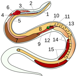
- Snakes: The esophagus is capable of extreme distension to accommodate large prey.
- Whales: Despite their size, a whale's esophagus is relatively narrow, generally measuring less than 10 cm in diameter (up to 25 cm in some baleen whales).
![1. Bile ducts: 2. Intrahepatic bile ducts 3. Left and right hepatic ducts 4. Common hepatic duct 5. Cystic duct 6. Common bile duct 7. Ampulla of Vater 8. Major duodenal papilla 9. Gallbladder 10–11. Right and left lobes of liver 12. Spleen 13. Esophagus 14. Stomach 15. Pancreas: 16. Accessory pancreatic duct 17. Pancreatic duct 18. Small intestine: 19. Duodenum 20. Jejunum 21–22. Right and left kidneys The front border of the liver has been lifted up (brown arrow).[42]](/images/dd/b5/ddb579d539dc1e634b83672d3264d889ff8d3877218d5ba5282768644fbdfdf3.webp)
Summary of Esophageal Characteristics
| Feature | Description/Detail |
|---|---|
| Tissue Type | Stratified squamous epithelium |
| Key Proteins | KRT13, KRT4, KRT6C (Keratins); MUC21, MUC22 (Mucins) |
| Primary Junction | Gastroesophageal junction (Z-line) |
| Common Diseases | Esophagitis, Barrett's esophagus, Esophageal cancer |
| Congenital Risks | Atresia and Fistula (1 in 3,500 births) |
Frequently Asked Questions
What is the z-line in the esophagus?
The z-line is the irregular, zig-zagging border at the gastroesophageal junction where the stratified squamous epithelium of the esophagus meets the simple columnar epithelium of the stomach.
Why can't horses vomit?
Horses cannot vomit because of a very well-developed muscular ring called the cardiac sphincter and the specific oblique angle at which the esophagus connects to the stomach.
What is the difference between esophageal atresia and a fistula?
Esophageal atresia is a condition where the esophagus ends in a blind sac rather than connecting to the stomach, whereas a fistula is an abnormal connection between the esophagus and the trachea.
What causes esophagitis?
Esophagitis can be caused by the reflux of stomach acid, infections (like yeast), allergic reactions, the ingestion of corrosive substances, or certain medications such as bisphosphonates.
How does the Nissen fundoplication work?
The Nissen fundoplication is a surgical procedure where the upper part of the stomach is wrapped around the lower esophageal sphincter to reinforce it and prevent gastric reflux.