Colloblasts: The Adhesive Hunting Mechanism of Ctenophores
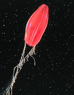
In the depths of the ocean, ctenophores—commonly known as comb jellies—utilize a sophisticated biological tool to secure their meals. While many are familiar with the stinging cells of jellyfish, tentaculate ctenophores employ a different strategy. They use colloblasts, unique multicellular structures designed for adhesion rather than envenomation.
Colloblasts serve as a synapomorphy (a shared derived characteristic) for tentaculate ctenophores, meaning they are a defining feature of this specific group. These specialized cells are distributed throughout the animal's tentacles, acting as a "sticky thicket" to immobilize prey upon contact.
Key Facts
- Function: Colloblasts use adhesives to stick to prey instead of using venom to sting.
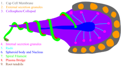
- Composition: They consist of a collocyte containing a spiral filament, internal granules, and other organelles.
- Distribution: Found in most ctenophores, except for the order Beroida and the genus Haeckelia.
- Mechanism: They are discharged from the tentilla when disturbed, releasing eosinophilic granules for adhesion.
Form and Function
First described in 1844, the colloblast is a complex cellular machine. It is primarily composed of two cell types: the stalk and the cap cells. The stalk is further divided into the collosphere (the head) and the collopod (the base). Surrounding the collosphere are numerous cap cells that deposit external secretion granules, also known as eosinophilic granules, which provide the necessary stickiness to capture prey.
A coiled spiral filament wraps around the collopod, which is anchored to the tentilla (the small branches of the tentacles) via a negatively charged root.
The Capture Process
Under normal conditions, the colloblast remains hidden within the pleated surface of the tentillum. The process of prey capture occurs in several rapid steps:
- Trigger: When the tentillum is disturbed, the plasma bridge connecting the spiral filament to the collopod breaks.
- Release: This break releases the colloblast from the tentilla.
- Adhesion: Upon contacting prey, the external secretion granules on the apical side of the collosphere rupture, releasing a powerful adhesive substance.
- Immobilization: Because the spiral filament remains attached to the tentillum, the prey is held firmly in place until it can be consumed.
Variation and Occurrence
While widespread, colloblasts are not universal across all comb jellies. They are absent in the order Beroida, which lacks tentacles entirely. Additionally, the genus Haeckelia does not produce its own colloblasts; instead, it utilizes cnidocytes (stinging cells) acquired from the cnidarian prey it consumes.
The most significant variation among groups is the shape of the collosphere. In the basal colloblast, the collosphere is spherical. However, in the orders Cestida, Cydippida, and Lobata, the collosphere is elongated and the collopod is absent. In these cases, the spiral filament attaches directly to the collosphere and is often reduced in size or missing entirely.
Polymorphism in Minictena luteola
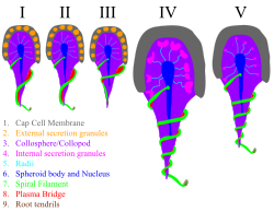
Within certain species, such as the cydippid ctenophore Minictena luteola, colloblasts exhibit polymorphism—meaning they exist in several different forms. These are categorized into five types based on the radii and the complexity of the spiral filament:
- Types I, II, and III: Located on the proximal side of the tentillum; these are smaller with fewer coils in their spiral filaments.
- Types IV and V: Located on the distal side; these feature more complex spiral filaments but lack external secretion granules.
Summary of Colloblast Characteristics
| Feature/Group | Basal Colloblast | Cestida, Cydippida, Lobata | Beroida / Haeckelia |
|---|---|---|---|
| Collosphere Shape | Spherical | Elongated | N/A |
| Collopod | Present | Absent | Absent |
| Spiral Filament | Attached to collopod | Attached to collosphere (or absent) | Absent |
| Capture Method | Adhesion | Adhesion | None / Stolen Cnidocytes |
Frequently Asked Questions
How do colloblasts differ from cnidocytes?
While both are used for prey capture, cnidocytes (found in cnidarians like jellyfish) are venomous stinging cells. Colloblasts, conversely, are non-venomous and use adhesive secretions to stick to their prey.
What are eosinophilic granules?
Eosinophilic granules are external secretion granules deposited by cap cells on the surface of the collosphere. They are the primary source of the adhesive substance used to capture prey.
Which ctenophores do not have colloblasts?
Colloblasts are absent in the order Beroida, which does not have tentacles, and in the genus Haeckelia, which uses cnidocytes obtained from its prey.
What happens when a colloblast is triggered?
When disturbed, a plasma bridge breaks, releasing the colloblast. Upon contact with prey, the granules on the collosphere rupture to release glue, while the spiral filament keeps the prey tethered to the animal.
What is the role of the spiral filament?
The spiral filament acts as a tether. It remains attached to the tentillum after the adhesive is released, ensuring that the captured prey cannot escape and is held until consumption.