Cellular Differentiation: The Mechanisms of Cell Specialization and Potency
Every complex multicellular organism begins as a single, simple cell called a zygote. The journey from this single cell to a sophisticated system of tissues and organs is driven by cellular differentiation (also known as cellular specification). This is the biological process by which a stem cell transforms into a more specialized cell type, altering its size, shape, metabolic activity, and responsiveness to external signals.
While differentiation is most prominent during embryonic development, it continues throughout adulthood. Adult stem cells facilitate tissue repair and normal cell turnover by dividing to create fully differentiated daughter cells. In some instances, differentiation is triggered by exposure to antigens.
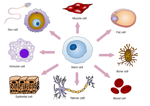
Key Facts
- Gene Expression: Differentiation is driven by controlled modifications in gene expression (epigenetics) rather than changes to the DNA sequence itself.
- Metabolic Shift: Stem cells possess abundant unsaturated metabolites, which decrease as the cell becomes more specialized.
- Potency: The ability of a cell to differentiate is termed "potency," ranging from totipotent (all cell types) to unipotent (one cell type).
- Reprogramming: Adult somatic cells can be reverted to a pluripotent state using specific transcription factors known as Yamanaka factors.
- Terminal Differentiation: Some cells permanently exit the cell cycle to perform a final, specialized function.
The Hierarchy of Cell Potency
Cell potency describes a cell's capacity to differentiate into other cell types. The higher the potency, the more diverse the range of cells it can produce.
- Totipotent: These cells can differentiate into all cell types, including placental tissue. In mammals, this is limited to the zygote and subsequent blastomeres.
- Pluripotent: These cells can become any cell type of the adult organism. Examples include embryonic stem cells in animals and meristematic cells in higher plants.
- Multipotent: These cells can differentiate into multiple, but closely related, cell types.
- Oligopotent: These are more restricted than multipotent cells, capable of becoming only a few closely related types.
- Unipotent: These cells can produce only one cell type but retain the ability for self-renewal.
In medical diagnostics, specifically cytopathology, the level of differentiation is used to determine the "grade" of a tumor, serving as a marker for cancer progression.
![Cell-count distribution featuring cellular differentiation for three types of cells (progenitor , osteoblast , and chondrocyte ) exposed to pro-osteoblast stimulus.[1]](/images/92/42/9242fdf943cf16143cf6f3bbcac96427c6b1cb43801b9588c3bd001e1461b29e.jpg)
Terminal Differentiation and Dedifferentiation
Terminal differentiation occurs when a precursor cell permanently leaves the cell cycle, dismantling its division machinery to focus entirely on its final function. This is critical in the vertebrate nervous system, the epidermis, the gut, and striated muscle. For example, a muscle cell will express specific genes for actin and myosin to facilitate contraction.
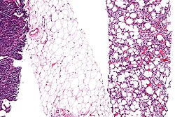
Conversely, dedifferentiation is the process where a specialized cell reverts to a simpler, less differentiated state. This is observed in certain contexts, such as amphibian limb regeneration or in the pathology of liposarcomas.
Mechanisms of Cellular Control
The transition from a stem cell to a specialized cell is governed by complex regulatory networks and signaling pathways.
Epigenetic Regulation
Epigenetics refers to modifications that affect gene expression without altering the DNA sequence. Key mechanisms include:
- DNA Methylation: The addition of methyl groups to DNA. Research shows that embryonic stem cells (ESCs) and induced pluripotent stem cells (iPSCs) share similar methylation levels (around 80% of CG dinucleotides), which are significantly higher than those in somatic cells (60%).
- Histone Modification: Changes to the proteins around which DNA is wrapped, affecting how accessible genes are for transcription.
- Nucleosome Positioning: The physical arrangement of DNA on nucleosomes that permits or restricts gene access.
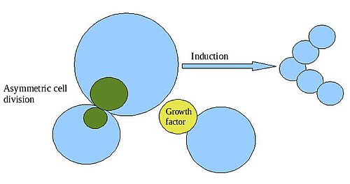
The Role of Pioneer Factors
Three critical transcription factors—OCT4, SOX2, and NANOG—are essential for maintaining pluripotency in embryonic stem cells. They manage chromatin structure to restrict or permit the transcription of target genes. The balance between these factors determines the cell's fate:
- High Oct4 / Low Sox2: Promotes a mesendodermal fate.
- High Sox2 / Low Oct4: Promotes a neural ectodermal fate.
- Nanog Suppression: A necessary prerequisite for any differentiation to occur.
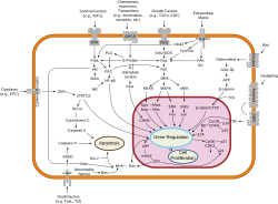
Induced Pluripotent Stem Cells (iPSCs)
Scientists can artificially revert adult somatic cells (like fibroblasts) back to a pluripotent state. This is achieved by introducing the Yamanaka factors: Oct4, Sox2, c-Myc, and Klf4. While iPSCs closely resemble ESCs in their genomic methylation patterns, they often retain some "epigenetic memory" from their original somatic progenitor cells.
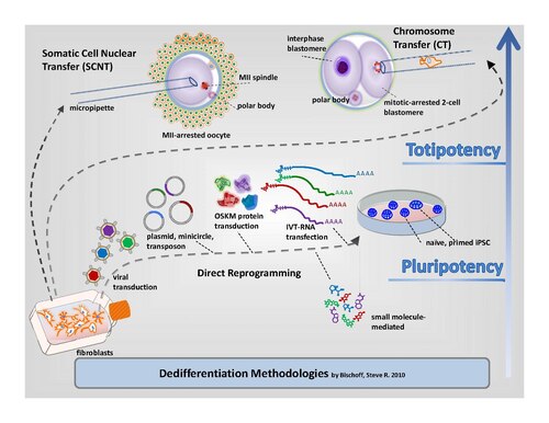
Summary of Cell Potency Levels
| Potency Level | Differentiation Capability | Example |
|---|---|---|
| Totipotent | All cell types + placental tissue | Zygote |
| Pluripotent | All adult organism cell types | Embryonic Stem Cells |
| Multipotent | Multiple closely related types | Adult Stem Cells |
| Oligopotent | A few closely related types | Lymphoid progenitor cells |
| Unipotent | Only one cell type | Skin basal cells |
Frequently Asked Questions
Does cellular differentiation change the DNA sequence?
No. With very few exceptions, differentiation does not change the DNA sequence. Instead, it relies on epigenetics—highly controlled modifications in gene expression that change which parts of the DNA are active.
What are Yamanaka factors?
Yamanaka factors are a group of four transcription factors (Oct4, Sox2, c-Myc, and Klf4) that can reprogram adult somatic cells, such as fibroblasts, back into a pluripotent state, creating induced pluripotent stem cells (iPSCs).
What is the difference between pluripotent and totipotent cells?
Totipotent cells can form every cell type in the body as well as extra-embryonic tissues like the placenta. Pluripotent cells can form any cell type within the adult body but cannot create a complete organism on their own because they cannot form placental tissue.
What happens during terminal differentiation?
During terminal differentiation, a cell permanently exits the cell cycle and stops dividing. It dismantles its division machinery and expresses genes specific to its final function, such as myosin in muscle cells.
How does DNA methylation differ between stem cells and somatic cells?
Embryonic and induced pluripotent stem cells typically show higher levels of CG dinucleotide methylation (around 80%) compared to somatic cells (around 60%). Additionally, pluripotent cells have higher levels of non-CG cytosine methylation (0.5% to 1.5%) than somatic cells.