Antibodies: The Precision Proteins of the Immune System
The human body possesses a sophisticated defense mechanism designed to identify and neutralize foreign invaders. At the heart of this system are antibodies, also known as immunoglobulins (Ig). These large proteins, part of the immunoglobulin superfamily, act as the immune system's targeted surveillance team, identifying and neutralizing antigens—substances such as bacteria and viruses that can trigger an immune response.
An antibody functions by recognizing specific antigens, which are essentially "antibody generators" of varying sizes and chemical compositions. This recognition is achieved through a high-precision binding mechanism where the antibody's paratope (the antigen-binding site) attaches to a specific epitope (a particular part of the antigen). This interaction is often compared to a lock and key, ensuring that each antibody targets only its intended match.
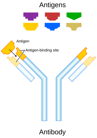
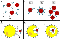
Once bound, antibodies can "tag" a microbe or infected cell for destruction by other immune cells or neutralize the threat directly—for instance, by blocking a virus from invading a host cell. While they are often discussed as secreted proteins floating in the extracellular space, they can also exist as B cell receptors anchored to the surface of immune cells.
Key Facts
- Dual Nature: Antibodies can be secreted freely into the blood or exist as receptors on B cells.
- High Specificity: Each antibody is designed to recognize specific epitopes on an antigen.
- Five Human Classes: Human antibodies are categorized into five isotypes: IgA, IgD, IgE, IgG, and IgM.
- Structural Diversity: The tips of the "Y" shape are highly variable to recognize millions of different antigens, while the stem is more constant.
- Effector Functions: The class of an antibody determines its function, where it is released, and its role in the immune response.
The Structure of an Antibody
![Angel of the West (2008) by Julian Voss-Andreae is a sculpture based on the antibody structure published by E. Padlan.[97] Created for the Florida campus of the Scripps Research Institute,[98] the antibody is placed into a ring referencing Leonardo da Vinci's Vitruvian Man thus highlighting the similarity of the antibody and the human body.[99]](/images/07/1e/071ed68eb5082f854111cd5e705a3378899d133f6cbd6b4ba15f87e10d5d7487.jpg)
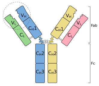
Structurally, an antibody is shaped like a "Y". It consists of four polypeptide chains: two identical heavy chains and two identical light chains. The branching arms of the "Y" contain the variable regions that form the paratopes, allowing the immune system to adapt to an almost infinite variety of pathogens.
The Antigen-Binding Site and Fc Region
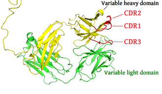
The tips of the antibody are the most variable parts, containing the complementarity determining regions that allow for precise antigen binding. In contrast, the stem of the "Y" is known as the Fc region. This region does not bind antigens but instead interacts with cell surface receptors (Fc receptors) and other proteins to trigger the actual immune response, such as phagocytosis (the ingestion of a pathogen by a phagocyte).
Antibody Complexes
Some antibodies are capable of binding multiple antigen molecules simultaneously, forming complexes. This is particularly evident in certain isotypes that can aggregate pathogens, making them easier for the immune system to clear.
Antibody Classes and Isotypes
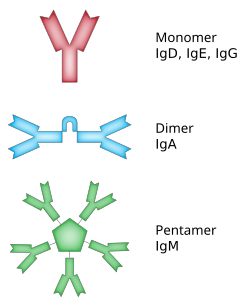
In humans, antibodies are divided into five main classes, or isotypes. The isotype determines the antibody's effector functions—the specific biological actions triggered after the antibody binds to its target.
| Class | Subclasses | Description/Role |
|---|---|---|
| IgA | 2 | Found in mucosal areas and secretions. |
| IgD | 1 | Primarily found on the surface of B cells. |
| IgE | 1 | Involved in allergic reactions and parasite defense. |
| IgG | 4 | Most abundant in blood; provides long-term immunity. |
| IgM | 1 | First antibody produced during an initial immune response. |
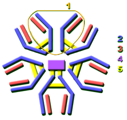
For example, secreted mammalian IgM is a pentamer, meaning it consists of five Ig units. With two binding sites per unit, a single IgM molecule can bind up to 10 epitopes, making it highly efficient at agglutination.
How the Body Generates Diversity
To defend against millions of different potential threats, the body uses complex genetic mechanisms to create a vast array of antibody shapes.
V(D)J Recombination
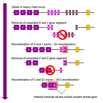
The primary method for creating diversity is V(D)J recombination. This process shuffles different gene segments (Variable, Diversity, and Joining) to create a unique sequence for the antigen-binding site of each B cell.
Somatic Hypermutation and Class Switching
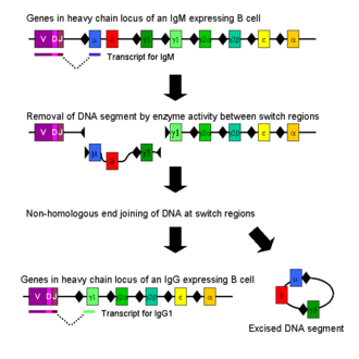
Once a B cell is activated, it can further refine its antibody through somatic hypermutation, which increases the binding affinity for the antigen. Additionally, class switch recombination allows an activated B cell to change the isotype of the antibody it produces (e.g., switching from IgM to IgG) to better suit the specific needs of the immune response.
Medical and Research Applications
The specificity of antibodies makes them invaluable tools in modern medicine and science. They are used in disease diagnosis to detect the presence of specific antigens (like viral proteins) or to identify if a patient has produced antibodies against a particular pathogen.
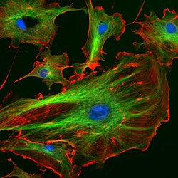
In research, antibodies conjugated to fluorescent molecules are used in immunofluorescence to visualize specific structures within cells, such as the cytoskeleton.
Beyond diagnostics, antibodies are used in therapy, including prenatal therapy and the development of monoclonal antibodies to treat various diseases. The production of these therapeutic antibodies requires rigorous validation to ensure purity and the elimination of viruses before clinical trials.
Frequently Asked Questions
What is the difference between an antibody and an immunoglobulin?
The terms are often used synonymously. However, "immunoglobulin" is a broader term that can refer to both the antibodies secreted into the blood and those that remain anchored to the surface of B cells as receptors.
How do antibodies actually "neutralize" a virus?
Antibodies can neutralize a virus by binding to the specific parts of the viral surface that the virus uses to attach to and enter a host cell, effectively blocking the entry point.
Why are there different classes of antibodies like IgG and IgM?
Different classes have different effector functions and distributions. For example, IgM is the first responder in an infection, while IgG provides long-term protection and can cross the placenta to protect a fetus.
What is an epitope?
An epitope is the specific part of an antigen that is recognized and bound by the antibody's paratope. A single antigen can have multiple different epitopes.
How does the body create so many different antibodies?
The body uses a genetic process called V(D)J recombination, which randomly combines different gene segments to create a nearly infinite variety of antigen-binding sites.