Opsonins and the Mechanisms of Immune Opsonization
In the complex landscape of the human immune system, the ability to identify and eliminate foreign invaders is critical for survival. One of the most effective ways the body achieves this is through opsonization—a process where specific molecules, known as opsonins, tag pathogens or dead cells to make them more "appetizing" and recognizable to phagocytic cells (cells that engulf and digest particles). This tagging system ensures that the immune response is both precise and efficient.
Opsonins are integral to both the innate immune system, which provides immediate, non-specific defense, and the adaptive immune system, which develops a tailored response to specific threats.
Key Facts
- Opsonins are molecules that mark antigens for destruction by phagocytes.
- The adaptive pathway primarily utilizes antibodies (IgG and IgM) and the complement system.
- The innate pathway uses complement proteins (C3b, C4b, iC3b) and lectins to recognize pathogens.
- PAMPs (Pathogen-Associated Molecular Patterns) are the specific molecular signatures that innate opsonins recognize.
- ADCC (Antibody-Dependent Cellular Cytotoxicity) allows NK cells to destroy tagged tumor or infected cells.
Adaptive Immune Opsonization
The adaptive immune response relies on antibodies, which are proteins synthesized by B cells. These antibodies are designed to bind to specific epitopes—unique regions on an antigen, such as a protein on a bacterial surface.
An antibody consists of two primary parts: the Fab region (antigen-binding region), which attaches to the epitope, and the Fc region (fragment crystallizable region). When the Fab region binds to a pathogen, the IgG antibody undergoes a conformational change. This change allows the Fc region to be recognized by Fc receptors (FcR) found on natural killer (NK) cells and other effector cells, triggering the release of lytic products to destroy the pathogen.
Beyond direct phagocytosis, antibodies can tag virally infected or tumor cells. NK cells then respond via the FcR in a process called antibody-dependent cellular cytotoxicity (ADCC).
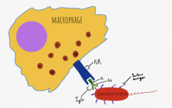
The Bridge to the Complement System
The adaptive response also recruits the complement system. When IgM or IgG antibodies bind to an antigen, they change shape, allowing the complement protein C1q to associate with the Fc region. This sequence eventually recruits C4b and C3b. These proteins are recognized by complement receptors 1, 3, and 4 (CR1, CR3, and CR4) located on most phagocytes, effectively bridging the adaptive and innate responses.
Additionally, C3d (a cleavage product of C3) recognizes PAMPs and opsonizes molecules to the CR2 receptor on B cells. This interaction lowers the threshold required for B cell activation via the B cell receptor, accelerating the adaptive response.
Innate Immune Opsonization
The innate immune system can opsonize pathogens immediately, often before the adaptive system has even been activated. This is primarily achieved through the complement system and specialized recognition proteins.
In the alternative pathway of complement activation, C3b is deposited directly onto antigens containing specific PAMPs, such as the lipopolysaccharides found on gram-negative bacteria. Once deposited, C3b is recognized by CR1 on phagocytes to initiate clearance.
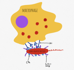
Another variant, iC3b, attaches to apoptotic cells and cellular remnants. By interacting with CR3 and CR4 on phagocytes, iC3b facilitates the removal of dead cells without triggering inflammatory pathways, ensuring a clean and quiet clearance process.
Lectins and Carbohydrate Recognition
The innate system also employs mannose-binding lectins (or ficolins), along with pentraxins and collectins. These molecules recognize specific carbohydrates expressed on the membranes of viruses, fungi, parasites, and bacteria. Once bound, they act as opsonins by activating phagocytic cells and the complement system.
Summary of Opsonization Components
| System | Primary Opsonins | Key Receptors | Target/Mechanism |
|---|---|---|---|
| Adaptive | IgG, IgM, C3d | FcR, CR2 | Specific epitopes; B cell activation |
| Innate | C3b, C4b, iC3b, Lectins | CR1, CR3, CR4 | PAMPs; apoptotic cells; carbohydrates |
Frequently Asked Questions
What is the difference between the Fab and Fc regions of an antibody?
The Fab region is the part of the antibody that binds to a specific epitope on an antigen. The Fc region is the constant fragment that is recognized by Fc receptors (FcR) on immune effector cells to initiate an attack.
How does iC3b differ from C3b in its function?
While C3b typically targets pathogens and triggers an immune response via CR1, iC3b attaches to apoptotic cells and bodies, facilitating their clearance via CR3 and CR4 without initiating inflammation.
What are PAMPs and why are they important for opsonization?
PAMPs, or Pathogen-Associated Molecular Patterns, are molecular signatures common to many pathogens (such as lipopolysaccharides on gram-negative bacteria). They allow innate opsonins like C3b to identify and tag invaders without needing prior exposure.
What is Antibody-Dependent Cellular Cytotoxicity (ADCC)?
ADCC is a mechanism where antibodies tag tumor cells or virally infected cells, which are then recognized and destroyed by natural killer (NK) cells via their Fc receptors.
How do lectins contribute to the opsonization process?
Mannose-binding lectins, ficolins, pentraxins, and collectins recognize specific carbohydrates on the membranes of bacteria, fungi, viruses, and parasites, subsequently activating phagocytic cells and the complement system.