Albert Einstein's Brain: Anatomy, Research, and the Odyssey of its Preservation
The mind of Albert Einstein changed the course of modern physics, but for decades, the physical organ that powered his genius was the subject of a strange and clandestine journey. Following his death in 1955, Einstein's brain became a focal point for scientists attempting to find a biological correlation between neuroanatomy and high-level mathematical intelligence.
Key Facts
- Weight: The brain was recorded at 1230 grams upon removal.
- Preservation: Removed by pathologist Thomas Stoltz Harvey, who kept it for over 20 years.
- Anatomical Anomalies: Research suggests a vacant parietal operculum and a thicker corpus callosum.
- Cellular Findings: Studies indicated a higher ratio of glial cells to neurons in the left inferior parietal area.
- Current Location: Remains are held by the National Museum of Health and Medicine and the Mütter Museum.
The Controversial Fate of the Brain
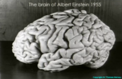
Shortly after Albert Einstein passed away in 1955, pathologist Thomas Stoltz Harvey performed the autopsy. During this process, Harvey removed the brain, weighed it, and later dissected it into approximately 240 blocks, each about 1 cm in size. To preserve these segments, he encased them in collodion, a plastic-like material, and used formalin for preservation.
The legality and ethics of this removal remain disputed. While some biographies suggest Einstein wanted his brain used for research, more recent evidence indicates the organ was taken without the explicit permission of Einstein or his immediate family. Although his son, Hans Albert Einstein, later endorsed the removal, he stipulated that any findings be published only in high-standing scientific journals.
For over two decades, the brain remained hidden in alcohol-filled mason jars inside a cider box. It was not until 1978 that journalist Steven Levy rediscovered the remains in Harvey's possession. Eventually, in 2010, Harvey's heirs transferred the holdings to the National Museum of Health and Medicine, while 46 small portions were acquired by the Mütter Museum in Philadelphia.
Scientific Analysis and Anatomical Findings
Researchers have spent years analyzing the preserved tissue and photographs to determine if Einstein's brain differed from the average human brain.
The Sylvian Fissure and Parietal Operculum
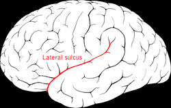
Analysis of autopsy photographs revealed an enlarged Sylvian fissure (also known as the lateral sulcus). A 1999 study by McMaster University found that the parietal operculum—a region in the inferior frontal gyrus of the frontal lobe—was vacant. Professor Sandra Witelson suggested that this unusual anatomy might have allowed neurons in that region to communicate more effectively, potentially explaining Einstein's preference for visual over verbal thinking.
Glial Cell Density
In the 1980s, Professor Marian Diamond conducted a study comparing Einstein's brain to 11 other male brains. She focused on glial cells, which are non-neuronal cells that provide support, nutrition, and myelin for signal transmission. Diamond found a statistically significant increase in the ratio of glial cells to neurons in the left inferior parietal area—a part of the association cortex responsible for synthesizing information from various brain regions.
The Hippocampus and Neocortex
In 2001, Dr. Dahlia Zaidel examined the hippocampus, a subcortical structure critical for learning and memory. She discovered that neurons on the left side of Einstein's hippocampus were significantly larger than those on the right. This asymmetry suggests stronger connections between the left hippocampus and the neocortex, the area where logical, analytical, and innovative thinking occurs.
Interhemispheric Communication
A 2013 study published in the journal Brain examined the corpus callosum, the thick bundle of fibers connecting the two cerebral hemispheres. Using high-resolution measurements, researchers found that Einstein's corpus callosum was thicker than those in both younger and older control groups, suggesting superior cooperation and communication between the two halves of his brain.
Summary of Anatomical Observations
| Brain Region/Feature | Observation in Einstein's Brain | Potential Significance |
|---|---|---|
| Parietal Operculum | Vacant/Absent | Enhanced neuronal communication |
| Glial Cells | Increased ratio (Left Inferior Parietal) | Higher metabolic support/synthesis |
| Left Hippocampus | Significantly larger neurons | Stronger link to the neocortex |
| Corpus Callosum | Increased thickness | Better interhemispheric communication |
| Mid-frontal Lobe | Fourth ridge present | Enhanced planning and working memory |
Critical Perspectives and Context
Despite these findings, some scientists urge caution. Critics point to publication bias, noting that studies showing differences are more likely to be published than those showing Einstein's brain was normal. Furthermore, because researchers knew which brain belonged to Einstein, conscious or unconscious bias may have influenced the results.
The practice of preserving the brains of geniuses is not unique to Einstein. Similar studies were conducted on the brain of mathematician Carl Friedrich Gauss, who had a brain weighing 1,492 grams with highly developed convolutions. Other preserved brains include those of Vladimir Lenin, Sofia Kovalevskaya, and the Native American Ishi.
Frequently Asked Questions
Was Einstein's brain larger than average?
No. His brain weighed 1230 grams, which is within the normal range. While some other historical figures, like Carl Friedrich Gauss or Edward H. Rulloff, had notably larger brains, Einstein's genius was not linked to overall size.
What are glial cells and why do they matter?
Glial cells are the support system of the brain; they provide nutrition, form myelin for faster signal transmission, and maintain the environment for neurons. A higher ratio of these cells in certain areas may indicate a more stimulating intellectual environment or higher efficiency in information processing.
Did Einstein consent to his brain being removed?
This is a matter of historical dispute. Some sources claim he insisted his brain be used for research, while more recent research suggests it was removed without the permission of Einstein or his close relatives.
What is the corpus callosum and why was Einstein's special?
The corpus callosum is the primary bridge of fibers connecting the left and right hemispheres of the brain. Einstein's was found to be thicker than average, which researchers believe may have facilitated better cooperation between the two hemispheres.
Where can the brain be seen today?
Portions of the brain, including thin slices on microscope slides, are part of the permanent galleries at the Mütter Museum in Philadelphia, while other remains are held by the National Museum of Health and Medicine.