Thoracic Vertebrae: Anatomy, Structure, and Regional Variations
The thoracic vertebrae form the middle segment of the vertebral column, situated between the cervical vertebrae of the neck and the lumbar vertebrae of the lower back. In humans, this region consists of twelve vertebrae, labeled T1 through T12. These bones serve as a critical structural bridge, increasing in size as they descend toward the lumbar region to support more of the body's weight.
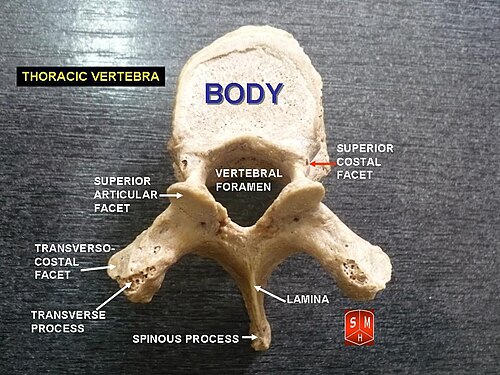
The defining characteristic of thoracic vertebrae is their relationship with the rib cage. They possess specialized facets—small, smooth articular surfaces—on the sides of their bodies and transverse processes, allowing them to articulate with the heads and tubercles of the ribs.
Key Facts
- Quantity: Humans have 12 thoracic vertebrae (T1–T12).
- Primary Function: Providing attachment points for the ribs and protecting the spinal cord.
- Unique Feature: Presence of costal demi-facets for rib articulation.
- Orientation: T1 is the uppermost thoracic vertebra, closest to the skull.
- Structural Trend: Vertebrae increase in size from T1 down to T12.
General Anatomical Characteristics
While each vertebra has unique traits, those from T2 to T8 share a consistent structural blueprint. The vertebral body in this middle region is typically heart-shaped and roughly equal in width and depth. These bodies are flat on the top and bottom, with a deep concavity at the back and a convex front.
A critical feature of these bodies is the presence of two costal demi-facets (half-facets) on each side. When the vertebrae are stacked, these demi-facets combine with the intervertebral fibrocartilage to create oval surfaces that receive the heads of the ribs.
The Vertebral Arch and Foramen
The posterior portion of the vertebra consists of the pedicles, which project backward and upward, and the laminae, which are broad, thick plates of bone. The laminae overlap the vertebrae below them, similar to tiles on a roof, to protect the spinal cord. Together, these structures form the vertebral foramen (or spinal canal), the central opening that houses the spinal cord.
Between each vertebral level are the intervertebral foramina. These small, circular openings allow the right and left nerve roots to exit the spinal canal.
Processes and Articulations
- Spinous Process: A long, triangular projection that points obliquely downward. From T5 to T8, these processes overlap.
- Transverse Processes: Strong, thick projections that extend backward and laterally. Most end in a concave surface for articulation with the rib tubercle.
- Articular Processes: The superior processes are thin plates projecting upward, while the inferior processes are fused largely with the laminae.
Individual Vertebral Variations
Certain thoracic vertebrae deviate from the general pattern to accommodate specific anatomical needs.
T1 and the Upper Region
The first thoracic vertebra (T1) resembles a cervical vertebra, featuring a broad, lipped body. Unlike others, it has a full articular facet for the first rib and a demi-facet for the second. Its spinous process is thick and nearly horizontal.
Mid-Thoracic Landmarks (T4–T9)
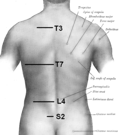
The T4 and T5 vertebrae align with the sternal angle. Notably, the human trachea typically divides into two main bronchi at the level of T5. Further down, T8 and T9 align with the xiphisternum (the lowest part of the sternum).
T10, T11, and T12: The Transition to Lumbar
As the spine transitions to the lumbar region, the vertebrae change significantly:
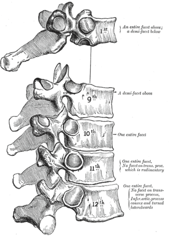
- T10: Features a full articular facet on each side rather than demi-facets and lacks facets on its lower surface.
- T11: The body increases in size to resemble lumbar vertebrae. The transverse processes are very short and lack articular facets.
- T12: Closely resembles a lumbar vertebra in its body, laminae, and spinous process. Its transverse process is divided into three tubercles (superior, inferior, and lateral).
Comparative Anatomy in Animals
The number of thoracic vertebrae varies widely across the animal kingdom. While humans have 12, most marsupials have 13, though koalas have only 11. Large mammals like horses, elephants, and rhinoceroses typically have 18 to 20. The extremes are found in certain sloths, which can have up to 25, and cetaceans (whales and dolphins), which may have as few as 9.
Summary of Thoracic Vertebrae
| Feature | T2–T8 (Typical) | T1 (Upper) | T11–T12 (Lower) |
|---|---|---|---|
| Body Shape | Heart-shaped | Broad, lipped (Cervical-like) | Large (Lumbar-like) |
| Rib Facets | Demi-facets | Full facet (1st rib) + demi-facet | Reduced or absent on transverse processes |
| Spinous Process | Long, oblique, overlapping | Thick, nearly horizontal | Short, nearly horizontal |
| Transverse Process | Long with rib facets | Long | Very short; T12 has three tubercles |
Frequently Asked Questions
How many thoracic vertebrae are in the human body?
There are twelve thoracic vertebrae, numbered T1 through T12.
What makes thoracic vertebrae different from cervical or lumbar vertebrae?
The primary distinction is the presence of costal facets on the bodies and transverse processes, which allow the thoracic vertebrae to articulate with the ribs.
What is the significance of the T4 and T5 levels?
T4 and T5 are located at the same level as the sternal angle, and the trachea typically bifurcates into the main bronchi at the level of T5.
Do all thoracic vertebrae connect to ribs in the same way?
No. While most use demi-facets, T1 has a full facet for the first rib, and T11 and T12 have shorter transverse processes that lack articular facets for rib tubercles.
Why are T11 and T12 different from the other thoracic vertebrae?
T11 and T12 serve as a transition zone, adopting characteristics of the lumbar vertebrae, such as larger bodies and different articular surface orientations, to support the increased weight of the upper body.