Sternum Anatomy: Structure, Development, and Clinical Significance
The sternum, commonly known as the breastbone, is a long, flat bone situated in the central part of the chest. Shaped roughly like a necktie, it serves as the anchor for the front of the rib cage by connecting to the ribs via cartilage. This structural arrangement is vital for protecting the heart, lungs, and major blood vessels from external injury. The term originates from the Ancient Greek word stérnon, meaning "chest."
Key Facts
- Composition: Consists of three primary regions: the manubrium, the body (gladiolus), and the xiphoid process.
- Function: Forms the anterior portion of the thoracic cage and protects vital organs.
- Landmark: The sternal angle is a critical clinical marker for locating the second rib.
- Development: Ossifies from six different centers, completing fusion typically by age 25.
- Clinical Use: Often used as a site for bone marrow biopsies, especially in patients with a high BMI.
Anatomical Structure
The sternum is a narrow, flat bone that angles obliquely downward and forward. It is slightly convex on the front and concave on the back. While it varies in size, it is generally longer in males than in females. The bone supports the clavicles (collarbones) at its top and attaches to the sternopericardial ligaments on its inner surface, which link the bone to the pericardium (the sac surrounding the heart).
The Manubrium
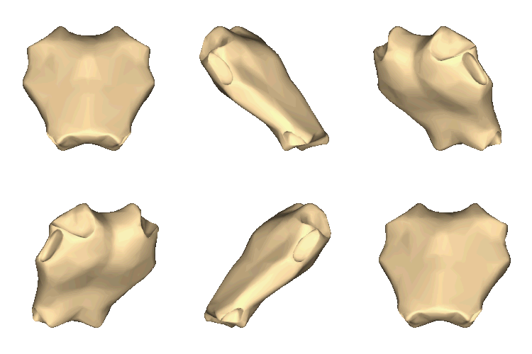
The manubrium is the broad, quadrangular superior portion of the sternum. Its most prominent feature is the suprasternal notch (or jugular notch), a palpable dip located between the two clavicles. The manubrium articulates with the clavicles and the costal cartilages of the first pair and a half of ribs.
The junction where the manubrium meets the body is known as the manubriosternal joint. This area forms the sternal angle, a palpable ridge that serves as a key anatomical landmark for healthcare providers.
The Body (Gladiolus)
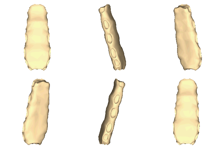
The body, or gladiolus, is the longest section of the sternum. It is characterized by three transverse ridges on its front surface, corresponding to the third, fourth, and fifth articular depressions. The pectoralis major muscle attaches to the sides of the body.
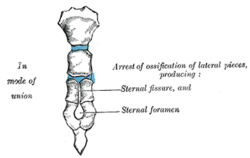
The lateral borders of the body contain depressions that receive the cartilages of the second through sixth ribs. The second rib specifically articulates at the sternal angle. In some individuals, a small opening called a sternal foramen may be present between the third and fourth segments.
The Xiphoid Process
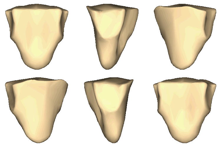
The xiphoid process is the small, pointed inferior extremity of the sternum. While it is the smallest part, it is clinically significant; during improperly performed cardiopulmonary resuscitation (CPR), the xiphoid process can snap off and potentially cause fatal hemorrhage by piercing the liver.
Development and Ossification
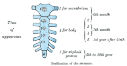
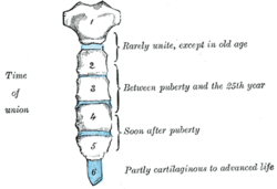
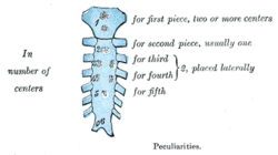
The sternum begins as two cartilaginous bars that fuse along the midline. It undergoes ossification (the process of cartilage turning into bone) from six distinct centers: one for the manubrium, four for the body (forming segments called sternebrae), and one for the xiphoid process.
Ossification begins during fetal life (sixth and seventh months) and continues after birth. The xiphoid process is the last to ossify, typically between the ages of five and eighteen. The segments of the body begin fusing around puberty, proceeding from the bottom upward, and are usually fully united by age 25. This process typically completes earlier in females than in males.
Clinical Significance and Variations
Surgical Procedures
In cardiothoracic surgery, a median sternotomy is performed, where the breastbone is cut open to provide access to the heart and lungs. The bone is later secured using wire cerclage or plates and screws. In rare cases of malignancy, the sternum may be totally removed (resected).
Trauma and Pathology
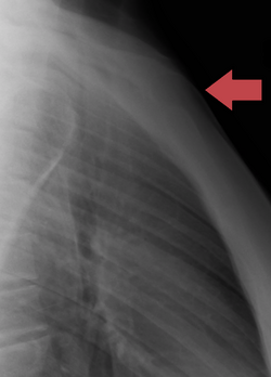
Sternal fractures are uncommon but can occur due to severe trauma, such as a chest hitting a steering column during a car accident. These are often comminuted fractures (broken into several pieces) and frequently occur at the sternal angle. They are often associated with pulmonary contusions (bruised lung tissue). Manubriosternal dislocation is rare and usually requires severe trauma or is facilitated by pre-existing arthritis.
Congenital Variations
- Sternal Foramen: A congenital hole present in 2.5% to 13.5% of the population, often mistaken for a bullet wound in forensic contexts.
- Bifid Sternum: An extremely rare failure of the sternal bars to fuse, resulting in a sternal cleft.
- Episternal Ossicles: Small bone fragments that may appear near the jugular notch.
Summary of Sternal Anatomy
| Region | Key Features | Primary Articulations |
|---|---|---|
| Manubrium | Suprasternal notch, Sternal angle | Clavicles, 1st & 2nd ribs |
| Body (Gladiolus) | Transverse ridges, Sternebrae | 2nd through 7th ribs |
| Xiphoid Process | Pointed inferior tip | 7th rib (partial) |
Frequently Asked Questions
What is the sternal angle and why is it important?
The sternal angle is the palpable ridge where the manubrium joins the body of the sternum. It is a critical clinical landmark because it marks the point where the second rib attaches, allowing clinicians to accurately count ribs and identify intercostal spaces during physical examinations.
Can a sternal foramen be dangerous?
A sternal foramen is generally asymptomatic and harmless. However, it can be problematic if a patient undergoes acupuncture in that specific area, as the needle could potentially pass through the hole into the thoracic cavity.
Why is the sternum used for bone marrow biopsies?
The sternum contains bone marrow and is easily accessible. It is particularly useful for patients with a high BMI (obese or overweight) who may have excess tissue making traditional biopsy sites, such as the pelvis, difficult to reach.
What happens during a median sternotomy?
A median sternotomy is a surgical procedure where the sternum is vertically divided to allow surgeons access to the heart and lungs. After the surgery, the bone is held together with surgical wires or plates to ensure proper healing and union.
How does the sternum develop in humans?
It develops from two cartilaginous bars that fuse in the center. It ossifies from six different centers—one in the manubrium, four in the body, and one in the xiphoid process—with full fusion typically occurring by age 25.