Solid Pseudopapillary Tumour of the Pancreas: Diagnosis and Pathology
A solid pseudopapillary tumour is a rare pancreatic neoplasm characterized by distinct morphological and immunohistochemical features. Identifying these tumours requires a precise diagnostic approach, combining advanced imaging-guided sampling with detailed microscopic analysis to differentiate them from other pancreatic masses.
Key Facts
- Gold Standard Diagnosis: Cytopathology via endoscopic ultrasound (EUS) guided fine needle aspiration (FNA).
- Typical Size: Ranges from 2 to 17 cm, with an average diameter of 8 cm.
- Defining Marker: Positive nuclear staining for beta catenin is found in 98% of cases.
- Morphology: Well-demarcated round masses containing both solid and cystic areas.
Diagnostic Procedures
The primary method for diagnosing these tumours is cytopathology—the study of individual cells—obtained through endoscopic ultrasound (EUS) guided fine needle aspiration (FNA). This minimally invasive procedure allows clinicians to extract a sample directly from the lesion for analysis.
Following the initial diagnosis, surgical excision is typically performed. Once the tumour is removed, it undergoes histopathology evaluation, which is the microscopic examination of tissue architecture, to determine the cancer stage.
Gross Morphology
Upon physical examination, solid pseudopapillary tumours typically appear as round, well-demarcated masses. These lesions generally measure between 2 and 17 cm in diameter, averaging around 8 cm. When cut sections are examined, the tumours exhibit a mixture of solid and cystic areas, often accompanied by hemorrhage.
Histomorphology and Cellular Structure
Under the microscope, these tumours are composed of solid sheets of cells that are focally dyscohesive, meaning the cells lack strong adhesion to one another. The cells typically feature uniform nuclei, occasional nuclear grooves, and cytoplasm that appears either clear or eosinophilic (pink-staining). A hallmark of these tumours is the presence of PAS-positive eosinophilic intracytoplasmic globules.
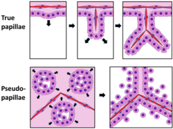
A critical feature of these tumours is the formation of pseudopapillae. Unlike true papillae, which are outgrowths of epithelium surrounding a fibrovascular core with a blood vessel, pseudopapillae form when cell death (necrosis) occurs in areas distant from blood vessels. This process leaves behind rims of cells lining the periphery of the nests, creating a deceptive papillary appearance.
Summary of Pathological Characteristics
| Feature | Observation |
|---|---|
| Average Diameter | 8 cm (Range: 2–17 cm) |
| Gross Appearance | Round, well-demarcated, solid/cystic with hemorrhage |
| Cellular Markers | PAS-positive intracytoplasmic globules |
| Nuclear Features | Uniform nuclei with occasional grooves |
Immunohistochemistry
Immunohistochemistry—the process of detecting specific proteins in cells using antibodies—is essential for confirming the diagnosis. Solid pseudopapillary tumours demonstrate positive nuclear staining for beta catenin, a finding present in 98% of cases.
Additional positive immunostaining is typically found for:
- CD10
- CD56
- Vimentin
- Alpha 1-antitrypsin
- Neuron specific enolase
Conversely, these tumours are negative for pancreatic enzymes and chromogranin, helping pathologists rule out other types of pancreatic neoplasms.
Frequently Asked Questions
What is the gold standard for diagnosing this tumour?
The gold standard is cytopathology performed via endoscopic ultrasound (EUS) guided fine needle aspiration (FNA) of the lesion.
How do pseudopapillae differ from true papillae?
True papillae are epithelial outgrowths with a central blood vessel. Pseudopapillae are formed when cells in the center of a nest disintegrate due to necrosis, leaving only the outer rims of cells.
Which protein marker is most characteristic of these tumours?
Beta catenin is the most characteristic marker, showing positive nuclear staining in 98% of solid pseudopapillary tumour cases.
What are the typical physical dimensions of these tumours?
They are generally round and well-demarcated, ranging from 2 to 17 cm in diameter, with an average size of 8 cm.
Are these tumours positive for pancreatic enzymes?
No, solid pseudopapillary tumours are negative for pancreatic enzymes and chromogranin.