Rhabdomyolysis: Diagnosis, Markers, and Potential Complications
Rhabdomyolysis is a serious clinical condition involving the breakdown of damaged skeletal muscle, which releases intracellular contents into the bloodstream. While it is often associated with acute trauma, crush injuries, or prolonged immobilization, it can also stem from underlying metabolic disorders. Timely diagnosis is critical, as the release of muscle proteins can lead to severe systemic complications, most notably acute kidney failure.
Key Facts
- Creatine Kinase (CK) is the most reliable diagnostic marker for rhabdomyolysis.
- CK levels above 1,000 U/L indicate rhabdomyolysis; levels over 5,000 U/L suggest severe disease.
- Kidney impairment is unlikely if CK levels remain below 20,000 U/L, absent other risk factors.
- Myoglobinuria (myoglobin in the urine) often causes a reddish-brown discoloration of the urine.
- Severe cases may lead to life-threatening complications like compartment syndrome or disseminated intravascular coagulation (DIC).
Diagnosing Rhabdomyolysis
Medical professionals may suspect rhabdomyolysis in patients with a history of physical trauma or those who have been immobilized for long periods. However, the condition is sometimes identified only after the onset of deteriorating kidney function, characterized by falling urine output and abnormally rising levels of urea and creatinine.
The Role of Creatine Kinase (CK)
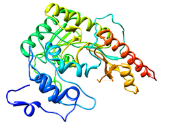
The gold standard for diagnosis is measuring the level of creatine kinase (CK), an enzyme released when muscle cells are damaged. CK levels typically rise steadily for 12 hours following the injury, remain elevated for one to three days, and then gradually decline.
There is a linear relationship between peak CK levels and the risk of acute kidney failure. While there is no absolute threshold for kidney damage, concentrations below 20,000 U/L generally do not cause impairment unless other risk factors are present. Mild elevations in CK without kidney impairment are termed hyperCKemia.
Other Biochemical Markers
While CK is the primary marker, other enzymes and proteins provide additional diagnostic clues:
- Myoglobin: This protein has a short half-life, making it less useful in later stages. However, its presence in blood or urine is linked to a higher risk of kidney impairment.
- Lactate Dehydrogenase (LDH) and Transaminases: These enzymes are often elevated. Because transaminases are found in both the liver and muscles, rhabdomyolysis can be mistaken for acute liver injury. Interestingly, actual acute liver injury occurs in 25% of non-traumatic rhabdomyolysis cases.
- Specialized Markers: Aldolase, troponin, carbonic anhydrase type 3, and fatty acid-binding protein (FABP) are typically reserved for diagnosing chronic muscle diseases.
Clinical Signs and Testing
Severe rhabdomyolysis often manifests through electrolyte imbalances. High potassium levels are common and can affect the heart's conduction system, which is detectable via electrocardiography (ECG) as T wave changes or a broadening of the QRS complex. Conversely, low calcium levels may appear initially as free calcium binds to damaged muscle cells.
Urine analysis often reveals a positive result for "blood" on test strips. This occurs because the reagent reacts with myoglobin, even when microscopy shows no actual red blood cells. This differs from hemolysis (the destruction of red blood cells), where the blood serum is visibly discolored. In cases of kidney damage, urine microscopy will show pigmented, granular urinary casts.
| Marker | Diagnostic Significance | Threshold/Observation |
|---|---|---|
| Creatine Kinase (CK) | Primary diagnostic tool | >1,000 U/L (Diagnosis); >5,000 U/L (Severe) |
| Myoglobin | Indicator of kidney risk | Causes reddish-brown urine; positive "blood" dipstick |
| Potassium | Severe case indicator | Elevated; may cause ECG changes |
| Calcium | Early stage indicator | Decreased due to muscle cell binding |
| Transaminases | General muscle/liver marker | Increased; can mimic liver injury |
Potential Complications
Compartment Syndrome
Compartment syndrome is a clinical diagnosis where pressure builds within a fascial compartment. Doctors assess severity by measuring the difference between the compartment pressure and the patient's blood pressure. A small difference indicates insufficient blood supply, which may necessitate surgical intervention.
Disseminated Intravascular Coagulation (DIC)
DIC is a critical illness complication characterized by unexpected bleeding or hematological abnormalities, such as a prolonged prothrombin time or a decreasing platelet count. This is typically confirmed using a D-dimer blood test.
Investigating Underlying Disorders
When rhabdomyolysis occurs without an obvious cause or happens repeatedly, further investigation into hereditary or metabolic muscle diseases is required.
- Metabolic Testing: Low blood carnitine and high acylcarnitine levels may suggest lipid metabolism defects. Disorders of glycolysis can be identified by measuring lactate after exercise; a failure of lactate to rise suggests a glycolysis disorder, while an exaggerated response suggests mitochondrial disease.
- Electromyography (EMG): This test can reveal specific patterns, such as the "cramp-like contracture" seen in phosphofructokinase deficiency or McArdle's disease.
- Muscle Biopsy: To avoid uninformative results caused by acute cell death, biopsies are usually delayed for several weeks. Histopathological analysis can identify specific markers, such as the "ragged red fibers" characteristic of mitochondrial diseases. MRI is often used to locate the most affected muscle sites for the biopsy.
Frequently Asked Questions
What is the most reliable test for rhabdomyolysis?
The most reliable test is the measurement of creatine kinase (CK) levels in the blood. Levels exceeding 1,000 U/L are indicative of rhabdomyolysis.
Why does a urine dipstick show blood when there are no red blood cells?
The reagent on the urine test strip reacts with myoglobin, a protein released from damaged muscles, which mimics the chemical signature of blood.
At what CK level does kidney damage become a concern?
While there is no absolute threshold, kidney impairment is generally unlikely if CK concentrations are below 20,000 U/L, provided no other risk factors are present.
Can rhabdomyolysis be mistaken for liver failure?
Yes, because transaminases are abundant in both muscle and liver tissue, their elevation during rhabdomyolysis can be confused with acute liver injury in the early stages.
What is the difference between hyperCKemia and rhabdomyolysis?
HyperCKemia refers to mild rises in creatine kinase levels that occur without associated kidney impairment.