Muscle Cells: Structure, Types, and Evolutionary Origins
Muscle cells, also known as myocytes, are specialized biological units designed for contraction and movement. These cells vary significantly in structure and function depending on their role in the body, ranging from the voluntary control of skeletal movement to the involuntary rhythmic beating of the heart and the regulation of internal organ diameter.
Key Facts
- Skeletal muscle fibers are multinucleated and formed by the fusion of embryonic myoblasts.
- Cardiac muscle cells are striated and feature a scalloped surface due to anchor fibers at the Z lines.
- Smooth muscle cells are spindle-shaped, lack sarcomeres, and are significantly shorter than skeletal fibers.
- Myogenesis is the process of muscle cell development, regulated by factors such as MyoD and Myf5.
- Striated muscles (skeletal and cardiac) contain organized units called sarcomeres, while smooth muscles do not.
Types of Muscle Cells
Skeletal Muscle Cells
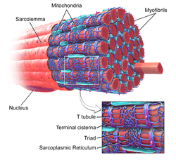
Skeletal muscle fibers are unique because they are multinucleated. This occurs during myogenesis (the formation of muscular tissue), where embryonic precursor cells called myoblasts fuse together to form a myotube. This fusion is facilitated by specific proteins known as fusogens, specifically myomaker and myomerger. To illustrate the scale of these structures, a single biceps brachii muscle in a young adult human male can contain approximately 253,000 muscle fibers.
Cardiac Muscle Cells
Found exclusively in the heart, cardiac muscle is striated, meaning it has a striped appearance under a microscope. Like skeletal muscle, it contains myofibrils (cylindrical bundles of proteins), myofilaments, and sarcomeres (the basic contractile units of muscle). A distinct feature of cardiac myocytes is their scalloped surface, created by transverse tubules emanating from grooves where anchor fibers (roughly 10 nm wide) secure the cell membrane to the cytoskeleton at the Z lines.
Smooth Muscle Cells
Smooth muscle cells differ fundamentally from striated muscles. They are spindle-shaped, featuring wide centers and tapering ends, with a single nucleus per cell. They are significantly smaller, ranging from 30 to 200 micrometers in length. Because of their small diameter, they do not require T-tubules. While they lack organized sarcomeres and myofibrils, they contain high concentrations of the contractile proteins actin and myosin. In these cells, actin filaments are anchored to the sarcolemma (cell membrane) by dense bodies, which serve a similar purpose to Z discs in striated muscle.
Muscle Cell Development
The development of all muscle types begins with the myoblast. The differentiation of these precursor cells is controlled by myogenic regulatory factors, including MyoD, Myf5, myogenin, and MRF4. Additionally, GATA4 and GATA6 are essential for the differentiation of cardiac myocytes, while the protein Kindlin-2 is necessary for the elongation of cells during myogenesis.
Comparative Summary of Muscle Cell Types
| Feature | Skeletal Muscle | Cardiac Muscle | Smooth Muscle |
|---|---|---|---|
| Appearance | Striated | Striated | Non-striated (Smooth) |
| Shape | Long Cylindrical | Branched/Scalloped | Spindle-shaped |
| Nuclei | Multinucleated | Single/Central | Single/Central |
| Sarcomeres | Present | Present | Absent |
| Control | Voluntary | Involuntary | Involuntary |
Evolutionary Perspectives
The origin of muscle cells is a subject of scientific debate. One theory, proposed by Schmid & Seipel (2005), suggests a monophyletic origin, meaning all muscle cells evolved from a single metazoan ancestor. They argue that similarities between the muscle cells of non-bilaterians (like Cnidaria and Ctenophora) and bilaterians indicate a common ancestor that was a triploblast (having three germ layers).
Conversely, Steinmetz, Kraus, et al. (2012) argue for a polyphyletic origin, suggesting that striated muscles evolved independently multiple times. They point out that key regulatory proteins, such as the troponin complex found in bilaterians, are missing in cnidarians and ctenophores. They also note that certain molecular markers, like myosin II, exist in sponges (Porifera) which lack true striated muscle, suggesting these proteins predated the evolution of muscle cells themselves.
Further research by Andrikou & Arnone (2015) utilizes gene regulatory networks to examine how tissue specification diverges among early deuterostomes and protostomes. Evidence suggests that specialized skeletal and cardiac muscles existed before the divergence of the vertebrate and arthropod lines, likely appearing more than 700 million years ago, while vertebrate smooth muscle evolved independently.
Frequently Asked Questions
What is the difference between a muscle fiber and a myofibril?
A muscle fiber is the entire muscle cell itself, whereas a myofibril is a smaller, protein-filled cylindrical structure found inside the muscle fiber.
Why do skeletal muscle cells have multiple nuclei?
Skeletal muscle cells are formed by the fusion of multiple individual myoblasts during development, with each myoblast contributing its own nucleus to the resulting cell.
Do smooth muscle cells have sarcomeres?
No, smooth muscle cells lack the organized sarcomeres and myofibrils found in striated muscle; instead, they use dense bodies to anchor their actin filaments.
What proteins are responsible for muscle contraction across all types?
All three types of muscle cells utilize the contractile proteins actin and myosin to generate force and movement.
How old are the earliest specialized muscle types?
Evidence indicates that specialized forms of skeletal and cardiac muscles developed in a common ancestor more than 700 million years ago.