Inner Ear Anatomy, Function, and Development
The inner ear (auris interna) is the innermost component of the vertebrate auditory system. While the outer and middle ear are primarily responsible for collecting and transmitting sound, the inner ear performs the critical tasks of sound detection and the maintenance of balance. In mammals, this complex system is housed within the temporal bone of the skull.
The inner ear is divided into two primary functional regions: the cochlea, which converts sound pressure into electrochemical impulses for the brain, and the vestibular system, which manages equilibrium and spatial orientation. Both systems are innervated by the eighth cranial nerve.
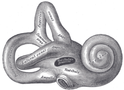
Key Facts
- Primary Functions: Responsible for hearing (auditory processing) and balance (vestibular processing).
- Core Structures: Comprises the bony labyrinth and the internal membranous labyrinth.
- Fluid Dynamics: Utilizes perilymph (outer spaces) and endolymph (inner spaces) to transmit signals.
- Sensory Mechanism: Uses specialized hair cells to detect mechanical vibrations and head movement.
- Blood Supply: The membranous labyrinth is supplied by the labyrinthine artery.
Structural Organization
The inner ear is organized into two concentric layers known as labyrinths.
Bony and Membranous Labyrinths
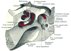
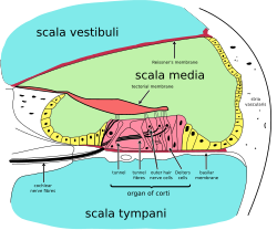
The bony labyrinth (osseous labyrinth) is a network of passages with bony walls lined with periosteum. It consists of three main parts: the vestibule, the semicircular canals, and the cochlea. Nested inside this is the membranous labyrinth, which creates three parallel fluid-filled spaces. The outer spaces are filled with perilymph, while the innermost space contains endolymph.
The Cochlear System and Hearing
Hearing begins when the middle ear's ossicles (malleus, incus, and stapes) translate pressure waves into mechanical vibrations. The stapes presses against the oval window, which is approximately 1/18 the area of the tympanic membrane, thereby increasing the pressure exerted on the perilymph fluid.
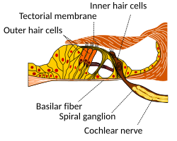
These fluid waves travel through the cochlea, where they are converted into nerve impulses. The cochlea's central axis, the modiolus, contains the spiral ganglion. The organ of Corti, located within the cochlear duct, is the actual sensory organ. It features a single row of inner hair cells and three rows of outer hair cells, which use actin-based stereocilia to detect vibrations.
The Vestibular System and Balance
The vestibular system detects the head's rotation, linear motion, and attitude. It works in tandem with the visual system and joint/muscle receptors to maintain balance. This system utilizes hair cells and mechanical structures such as the curved semicircular canals and calcium carbonate crystals called otoliths found in the saccule and utricle.
Microanatomy of the Organ of Corti
The organ of Corti is a highly specialized structure containing various supporting cells that maintain the environment for sensory hair cells:
- Pillar Cells: Provide mechanical coupling between the basement membrane and mechanoreceptors.
- Deiters' Cells: Neuroglial cells that support the hair cell area.
- Claudius' and Boettcher's Cells: Supporting cells involved in ion transport and sealing endolymphatic spaces.
- Hensen's Cells: High columnar cells adjacent to Deiters' cells.
Development and Blood Supply
In humans, the inner ear begins developing in the fourth week of embryonic growth from the auditory placode (a thickening of the ectoderm). This forms the otocyst (auditory vesicle), which eventually differentiates into the saccule, utricle, and the cochlear duct by the fifth week.
The blood supply to the bony labyrinth comes from the anterior tympanic, petrosal, and stylomastoid branches. The membranous labyrinth is specifically supplied by the labyrinthine artery, with drainage occurring via the labyrinthine vein into the sigmoid or inferior petrosal sinus.
Comparative Anatomy Across Species
| Group | Cochlea Presence | Key Features |
|---|---|---|
| Mammals | Yes (Coiled) | Complex organ of Corti; coiled cochlea for space efficiency. |
| Birds/Crocodilians | Yes (Straight) | Elongated, slightly curved bony tube. |
| Reptiles/Amphibians | No (Lagena) | Use a lagena or papilla amphibiorum for sound detection. |
| Fish | No | Sound transmitted via skull bones or swim bladder; use macula neglecta. |
Clinical Disorders
Interference with the labyrinth can lead to labyrinthitis, an inflammation caused by infection or blockage. Symptoms include vertigo, nausea, and disorientation. Another rare condition is autoimmune inner ear disease (AIED), which causes rapidly progressive, bilateral sensorineural hearing loss.
Frequently Asked Questions
What is the difference between perilymph and endolymph?
Perilymph is the fluid that fills the two outer spaces of the membranous labyrinth, while endolymph is the fluid contained within the innermost membranous space.
How does the inner ear detect balance?
The inner ear uses the semicircular canals to detect rotational movement and the saccule and utricle (containing otoliths) to detect linear acceleration and gravity.
What is the role of the organ of Corti?
The organ of Corti is the sensory organ of hearing; it contains hair cells that convert mechanical fluid vibrations into electrical signals for the auditory nerve.
What causes the sensation of vertigo in labyrinthitis?
Vertigo occurs when inflammation or infection in the labyrinth disrupts the vestibular system's ability to accurately send balance and motion data to the brain.
How does hearing frequency vary in the cochlea?
High-frequency sounds cause the base of the basilar membrane to move the most, while low-frequency sounds cause the tip (apex) of the membrane to move the most.