Image Guided Planning in Radiotherapy
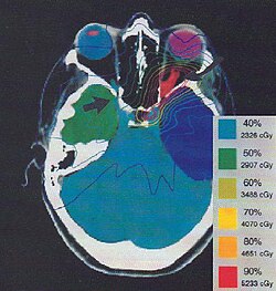
Modern radiotherapy relies on a sophisticated process known as image guided planning to ensure that radiation is delivered with extreme precision. By creating a virtual patient through medical imaging, clinicians can design a treatment strategy that maximizes the dose to the tumor while minimizing exposure to surrounding healthy tissues.
The Foundation of Treatment Planning
The process begins with the acquisition of high-quality medical images to form a digital model of the patient. A CT scan (Computed Tomography) typically serves as the primary image set. To enhance the accuracy of soft tissue contouring—the process of outlining organs and tumors—MRI (Magnetic Resonance Imaging) is often used as a secondary set. In specific cases, PET scans (Positron Emission Tomography) are utilized when uptake studies are needed to better define the planning target volume.
To combine these different imaging sources, modern systems use multimodality image matching, also referred to as image coregistration or fusion. This allows clinicians to overlay different types of scans for a comprehensive view of the patient's anatomy.
Treatment simulations then address the geometric, radiological, and dosimetric aspects of the therapy. For Intensity Modulated Radiation Therapy (IMRT), this involves selecting the beam type (photons, electrons, or protons), the energy level (such as 6 or 18 megaelectronvolt (MeV) photons), and the physical arrangement of the equipment.
In the case of brachytherapy—where radiation sources are placed inside the body—planning focuses on catheter positions and source dwell times for High Dose Rate (HDR) therapy, or the specific placement of seeds for Low Dose Rate (LDR) therapy.
Key Facts
- Primary Imaging: CT scans are the standard for primary treatment planning.
- Secondary Imaging: MRI is preferred for soft tissue contouring; PET is used for specific uptake studies.
- Optimization: Plans are evaluated using dose-volume histograms to check dose uniformity and organ sparing.
- Planning Methods: Radiotherapy uses two primary optimization strategies: forward planning and inverse planning.
- Calculation Models: Dose distribution is calculated using pencil beam, convolution-superposition, or Monte Carlo simulations.
Forward Planning
Forward planning is a manual process where the planner determines the beam parameters to achieve the desired dose. The planner must decide on the number of beams, the angles of delivery, the use of attenuating wedges, and the MLC (Multi-Leaf Collimator) configuration used to shape the radiation beam.
Once the initial plan is set, the system calculates the required monitor units to deliver the prescribed dose. This distribution depends on the patient's anatomy and beam modifiers, such as field size and tumor depth. To model how radiation travels through tissue, planners use various calculation models, balancing precision against computation time via pencil beam, convolution-superposition, or Monte Carlo simulations.
Because of its manual nature, forward planning is generally reserved for simpler cases where the tumor has a basic shape and is located away from critical organs.
Inverse Planning
Inverse planning reverses the manual trial-and-error approach. In this method, the radiation oncologist defines the tumor and critical organs, and the planner assigns target doses and importance factors for each area.
An optimization program then solves the "Inverse Problem," automatically calculating the treatment plan that best matches the input criteria. This allows for much more complex dose distributions than forward planning can typically achieve.
| Feature | Forward Planning | Inverse Planning |
|---|---|---|
| Process | Manual selection of beams and angles | Optimization based on target goals |
| Workflow | Trial-and-error adjustment | Algorithmic solving of the Inverse Problem |
| Complexity | Best for simple tumor shapes | Ideal for complex shapes near critical organs |
| Input | Beam parameters and modifiers | Target doses and importance factors |
Frequently Asked Questions
What is the difference between forward and inverse planning?
Forward planning involves the planner manually choosing beam angles and modifiers to reach a dose goal. Inverse planning involves setting the desired dose goals first and letting a computer program calculate the best beam configuration to achieve them.
Why are multiple types of scans used in planning?
Different scans provide different information: CT scans are primary for planning, MRI provides superior detail for soft tissues, and PET scans help identify specific metabolic activity to better define the target volume.
What are dose-volume histograms?
Dose-volume histograms are tools used by clinicians to evaluate a plan's quality, specifically checking if the tumor receives a uniform dose and ensuring that healthy surrounding structures are sufficiently spared.
What is image coregistration?
Image coregistration, or fusion, is the process of matching and overlaying multiple imaging modalities (like CT and MRI) into a single coordinate system to improve the accuracy of treatment planning.
What are the common dose calculation models used?
The most common models are pencil beam, convolution-superposition, and Monte Carlo simulation, with the choice depending on the required balance between calculation precision and the time needed for computation.