Hodgkin Lymphoma: Diagnosis, Types, and Staging
Hodgkin lymphoma is a malignancy of the lymphatic system that requires precise diagnostic techniques to distinguish it from noncancerous lymph node swelling, such as infections, and other forms of cancer. The process of identifying this disease involves a combination of tissue analysis, advanced imaging, and systemic evaluations to determine the extent of the disease and the most appropriate treatment path.
Key Facts
- Definitive Diagnosis: Confirmed via lymph node biopsy, typically an excisional biopsy with microscopic examination.
- Primary Markers: The presence of Reed-Sternberg cells is a hallmark of the disease.
- Main Types: Divided into Classic Hodgkin Lymphoma (approx. 90%) and Nodular Lymphocyte-Predominant Hodgkin Lymphoma (approx. 10%).
- Imaging: PET scans are the gold standard for detecting small deposits and functional imaging of high-metabolism tissues.
- Staging: The Ann Arbor system is commonly used to classify the spread of the disease across the diaphragm.
Diagnostic Procedures
To reach a definitive diagnosis, clinicians perform a lymph node biopsy to examine the tissue microscopically. In addition to biopsy, blood tests are conducted to evaluate major organ function and ensure the patient can safely undergo chemotherapy.
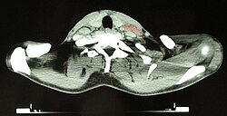
Imaging plays a critical role in detecting the disease. While CT scans are common, Positron Emission Tomography (PET) is used to find small deposits that CT might miss. PET scans utilize radiolabeled glucose to create functional images of tissues with high metabolic activity. In specific instances, a gallium scan may be used as an alternative to PET.
Types of Hodgkin Lymphoma
Hodgkin lymphoma is categorized into two primary types based on morphology, phenotype, and molecular features, which influence how the disease behaves clinically.
Classic Hodgkin Lymphoma
Accounting for roughly 90% of cases, classic Hodgkin lymphoma is further divided into four pathologic subtypes. These subtypes are defined by the morphology of the Reed-Sternberg cells (malignant cells) and the composition of the reactive cell infiltrate (the surrounding immune cells).
| Subtype | Description | ICD-10 | ICD-O |
|---|---|---|---|
| Nodular sclerosing HL | Most common; features large tumor nodules, lacunar RS cells, and collagen fibrosis/sclerosis. | C81.1 | M9663/3 |
| Mixed-cellularity HL | Common; numerous RS cells mixed with inflammatory cells; often associated with Epstein-Barr virus (EBV) and immunocompromised states. | C81.2 | M9652/3 |
| Lymphocyte-rich HL | Rare; resembles B-cell non-Hodgkin lymphoma; carries the most favorable prognosis. | C81.0 | M9651/3 |
| Lymphocyte depleted HL | Rare; characterized by many pleomorphic RS cells and few reactive lymphocytes. | C81.3 | M9653/3 |
The Epstein-Barr virus (EBV) is frequently found in the Reed-Sternberg cells of the lymphocyte-depleted (>70%) and mixed-cellularity (70%) subtypes, and is present in about 40% of lymphocyte-rich cases.
Nodular Lymphocyte-Predominant Hodgkin Lymphoma (NLPHL)
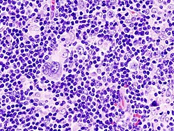
Representing about 10% of cases, NLPHL is distinct from the classic form. It is characterized by popcorn cells, which express the CD20 marker. Because of these biological differences, NLPHL is often treated differently, sometimes utilizing rituximab in combination with AVBD chemotherapy.
Pathology and Microscopy
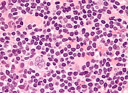
Macroscopically, affected lymph nodes (often in the laterocervical region) are enlarged but maintain their shape because the capsule is not invaded. The cut surface is typically uniform white-grey, though nodular sclerosis may present a nodular appearance.
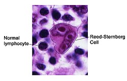
Under a microscope, the lymph node architecture is partially or completely replaced by scattered malignant Reed-Sternberg cells (RSC). These are large, often bi-nucleated cells (20–50 micrometres) with prominent nucleoli, often described as having "owl eyes" due to mirror-image nuclei.
Reed-Sternberg Cell Variants
- Hodgkin cell: A mononucleated version of the RS cell.
- Lacunar RSC: Features a single hyperlobulated nucleus with cytoplasm retracted to create empty spaces called "lacunae."
- Pleomorphic RSC: Contains multiple irregular nuclei.
- Popcorn RSC: A smaller cell with a highly lobulated nucleus and small nucleoli.
- Mummy RSC: Features a compact nucleus without a nucleolus.
Staging the Disease
Staging determines which areas of the body are affected through physical exams, blood tests, chest X-rays, CT, PET, or MRI scans, and usually a bone marrow biopsy. The Ann Arbor staging classification is the standard scheme used:
- Stage I: Involvement of a single lymph node region or a single extralymphatic site.
- Stage II: Involvement of two or more lymph node regions on the same side of the diaphragm, or one region and a contiguous extralymphatic site.
- Stage III: Involvement of lymph node regions on both sides of the diaphragm, including the spleen or limited contiguous extralymphatic sites.
- Stage IV: Disseminated involvement of one or more extralymphatic organs.
Additional modifiers are added to the stage: "A" for the absence of systemic symptoms, "B" for the presence of symptoms, "E" for localized extranodal extension, "S" for splenic involvement, and "X" for bulky disease.
Frequently Asked Questions
How is Hodgkin lymphoma definitively diagnosed?
The definitive diagnosis is made through a lymph node biopsy, typically an excisional biopsy, followed by a microscopic examination to identify malignant Reed-Sternberg cells.
What is the difference between classic HL and NLPHL?
Classic Hodgkin lymphoma (90% of cases) is characterized by classic Reed-Sternberg cells and divided into four subtypes. Nodular lymphocyte-predominant Hodgkin lymphoma (10% of cases) is characterized by "popcorn cells" that express CD20 and often requires different treatment strategies.
What are Reed-Sternberg cells?
Reed-Sternberg cells are large, malignant cells found in the lymph nodes of patients with Hodgkin lymphoma. They are typically bi-nucleated with prominent nucleoli, giving them an "owl eye" appearance under a microscope.
What does the Ann Arbor staging system measure?
The Ann Arbor system classifies the extent of the lymphoma based on whether the disease is limited to one lymph node region, spread to multiple regions on one side of the diaphragm, spread to both sides of the diaphragm, or disseminated to extralymphatic organs.
Why is a PET scan used instead of just a CT scan?
PET scans are more sensitive for detecting small deposits of lymphoma and provide functional imaging by identifying tissues with high glucose metabolism, which is characteristic of malignant cells.