Depolarization: The Electrical Engine of Cellular Communication
In the complex world of biology, communication happens at lightning speed through electrical shifts. At the heart of this process is depolarization (also known as hypopolarization), a change in a cell's electric charge distribution that makes the interior of the cell less negative compared to its exterior. This mechanism is fundamental to how neurons fire, how muscles contract, and how our organs coordinate their functions.
While most cells maintain a negative internal charge relative to the outside—a state known as the membrane potential—depolarization temporarily shifts this balance. In some cases, such as during an action potential, the charge reverses entirely, leaving the inside of the cell positively charged. This shift is typically driven by an influx of cations (positively charged ions), most commonly sodium, or an efflux of anions (negatively charged ions).
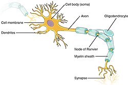
Key Facts
- Definition: A shift in membrane potential that makes the cell interior less negative.
- Primary Driver: The influx of sodium ions (Na+) into the cell.
- Essential Role: Critical for electrical signaling in neurons and muscle cells.
- Opposite Process: Hyperpolarization, where the cell becomes more negative than its resting state.
- Key Protein: The sodium-potassium pump maintains the gradients necessary for depolarization to occur.
The Foundation: Establishing the Resting Potential
Before a cell can depolarize, it must first establish a resting potential. This is a state of electrical imbalance where the interior of the cell is negatively charged. This environment is created and maintained by specialized transmembrane proteins embedded in the plasma membrane.
The Role of the Sodium-Potassium Pump
The sodium-potassium pump is an ATPase (an enzyme that uses ATP for energy) that actively transports three sodium ions (Na+) out of the cell for every two potassium ions (K+) it brings in. This process achieves two things: it makes the interior less positive and creates a steep concentration gradient, with high sodium levels outside and high potassium levels inside.
Additionally, potassium leak channels allow a controlled passive exit of potassium ions, and internal negatively charged components further stabilize the negative inner charge. This setup ensures that the cell is "primed" and ready to fire when a stimulus arrives.
The Mechanics of Depolarization and Action Potentials
Once the resting potential is set, the cell can undergo depolarization. This process is the catalyst for the action potential, the rapid electrical impulse that travels along a cell membrane.
The Depolarization Phase
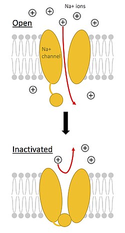
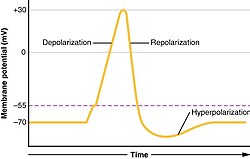
When a cell is stimulated, voltage-gated sodium and calcium channels open. Sodium ions rush into the cell, following their electrochemical gradient. This influx adds positive charge to the interior, shifting the membrane potential from negative toward positive. This creates a positive-feedback loop: the initial influx of sodium triggers even more sodium channels to open, sustaining the process until a positive equilibrium potential is reached.
Repolarization and Hyperpolarization
To prevent the cell from remaining permanently positive, sodium channels possess an inherent inactivation mechanism that closes them rapidly. Once the interior is sufficiently positive, voltage-gated potassium channels open. Potassium ions move out of the cell, decreasing the internal potential and returning it toward the resting state—a process called repolarization.
Often, this process overshoots the mark. So many potassium ions leave the cell that the potential becomes more negative than the original resting potential. This state is known as hyperpolarization. Eventually, the sodium-potassium pump and the closing of ion channels restore the cell to its baseline resting potential.
Depolarization in Action: Neurons, Eyes, and the Heart
Neural Transmission
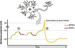
In neurons, depolarization is the primary method of transmitting stimuli. Stimuli can be excitatory (increasing voltage and making depolarization easier) or inhibitory (causing hyperpolarization, which makes the cell harder to depolarize). These signals converge at the axon hillock, where they are summed. If the total voltage reaches the threshold potential, an action potential surges down the axon to the terminal, triggering the release of neurotransmitters.
The Unique Case of Rod Cells
In the rod cells of the eye, depolarization works differently. In the dark, rod cells are naturally depolarized, constantly releasing neurotransmitters. When light is absorbed, the sodium and calcium channels close, causing the cell to hyperpolarize and release fewer neurotransmitters. In this specific instance, depolarization prevents a signal from reaching the brain, while hyperpolarization initiates it.
Cardiac Coordination
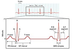
The heart relies on a precise sequence of depolarization to pump blood. The sinoatrial (SA) node initiates depolarization in the atria, causing them to contract. This wave then reaches the atrioventricular (AV) node, which introduces a 100 ms delay before triggering the depolarization and contraction of the ventricles.
Summary of the Electrical Cycle
| Stage | Primary Action | Ion Movement | Effect on Potential |
|---|---|---|---|
| Trigger | Excitatory signal | Initial shift | Less negative |
| Upstroke | Voltage-gated Na+ channels open | Sodium influx | Rapid depolarization |
| Peak | Na+ channels inactivate | Sodium influx stops | Positive equilibrium |
| Repolarization | Voltage-gated K+ channels open | Potassium efflux | Returns toward rest |
| Hyperpolarization | K+ channels remain open briefly | Excess potassium efflux | More negative than rest |
Depolarization Blockers
Certain pharmacological agents, known as depolarization blocking agents, can interfere with this cycle. Drugs like suxamethonium (succinylcholine) and decamethonium act as nicotinic agonists. They keep the cell membrane depolarized by preventing channels from closing. Because the membrane cannot repolarize, the voltage-gated sodium channels remain inactivated, effectively blocking further electrical transmission.
Frequently Asked Questions
What is the difference between depolarization and hyperpolarization?
Depolarization occurs when the cell's internal charge becomes less negative (more positive), moving it closer to the threshold for firing an action potential. Hyperpolarization occurs when the internal charge becomes more negative than the resting potential, making it harder for the cell to fire.
How does the sodium-potassium pump contribute to depolarization?
While the pump itself creates a negative charge, its primary role is to establish the concentration gradients (high sodium outside, high potassium inside) that allow ions to rush into or out of the cell rapidly once the voltage-gated channels open.
What happens during the recovery period of a neuron?
After an action potential, the neuron must restore its resting membrane potential. During this recovery period, the neuron is unable to transmit another action potential until the electrical balance is reset.
How is depolarization represented on an ECG?
On an electrocardiogram, the P wave represents atrial depolarization, and the QRS complex represents ventricular depolarization. The T wave represents the subsequent ventricular repolarization.
Why do rod cells in the eye behave differently than neurons?
Unlike most neurons where depolarization triggers a signal, rod cells are depolarized in the dark and release neurotransmitters constantly. Light causes them to hyperpolarize, which reduces neurotransmitter release and signals the brain that light has been detected.