Cell Nucleus: Structure, Function, and Nuclear Architecture
The nucleus serves as the command center of the eukaryotic cell, acting as the repository for genetic information and the coordinator of cellular activities. By sequestering the cell's DNA, the nucleus allows for sophisticated regulation of gene expression and protects the genome from metabolic byproducts in the cytoplasm.
From the intricate network of the nuclear envelope to the specialized subnuclear bodies, the architecture of the nucleus is designed for efficiency, ensuring that replication and transcription occur in a highly organized environment.
Key Facts
- Genetic Storage: The nucleus houses the majority of a cell's DNA, complexed as chromatin.
- Selective Permeability: The nuclear envelope regulates the movement of macromolecules via specialized nuclear pores.
- Ribosome Production: The nucleolus is the dedicated site for ribosome synthesis.
- Structural Support: The nuclear lamina provides mechanical stability to the nuclear envelope.
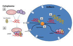
- Dynamic Organization: The nucleus contains various non-membrane-bound bodies, such as Cajal bodies and speckles, to facilitate specific biochemical reactions.
Nuclear Structure and Landmarks
The Nuclear Envelope and Pores
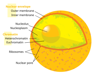
The nucleus is enclosed by the nuclear envelope, a double-membrane system. The outer membrane is continuous with the rough endoplasmic reticulum and is often studded with ribosomes. This envelope is perforated by nuclear pores, complex protein channels that control the traffic of RNA and proteins between the nucleus and the cytosol.
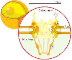
A cross-section of a nuclear pore reveals a sophisticated structure consisting of an outer ring, spokes, a nuclear basket, and cytoplasmic filaments. These components ensure that only authorized macromolecules enter or exit the nucleus.
The Nuclear Lamina
Just inside the inner nuclear membrane lies the nuclear lamina, a dense fibrillar network of intermediate filaments called lamins. The lamina provides structural support, maintains the shape of the nucleus, and plays a critical role in organizing chromatin and regulating DNA replication and transcription.
The Nucleolus
The nucleolus is the most prominent subnuclear structure. It is a non-membrane-bound region where ribosomal RNA (rRNA) is synthesized and combined with proteins to form ribosome subunits.
Chromosomes and Genetic Organization
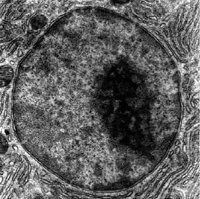
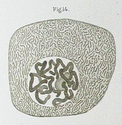
Inside the nucleus, DNA is not randomly distributed. It is complexed with proteins to form chromatin. During interphase, chromatin is relatively loose, but during mitosis, it condenses into distinct chromosomes.
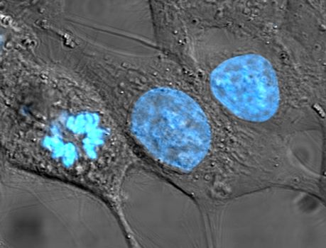
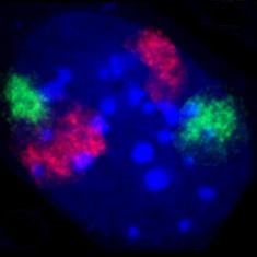
Research indicates that chromosomes occupy specific chromosome territories rather than being entangled. Active and inactive genes often localize preferentially toward the periphery of these territories to optimize access for transcription machinery.
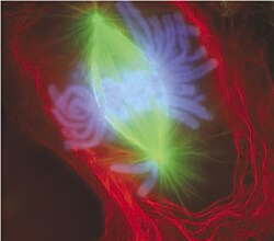
During cell division (metaphase), chromosomes align at the metaphase plate, where they are attached to the mitotic spindle for equal distribution into daughter cells.
Subnuclear Bodies and Compartmentalization
The nucleus utilizes compartmentalization to increase the efficiency of gene expression. Various nuclear bodies act as hubs for specific molecular processes.
Specialized Nuclear Bodies
- Cajal Bodies: Involved in the assembly of small nuclear ribonucleoproteins (snRNPs).
- Splicing Speckles: Regions rich in splicing factors that assist in the processing of pre-mRNA.
- PML Bodies: Involved in various cellular responses, including apoptosis and antiviral defense.
- Paraspeckles: Built on long non-coding RNA and involved in gene regulation.
Transcription Factories
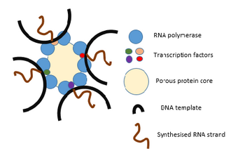
Transcription often occurs in transcription factories—protein-rich cores where multiple RNA polymerases can transcribe several genes simultaneously, regardless of their original position on the chromosome.
Nuclear Function and Dynamics
Nuclear Transport
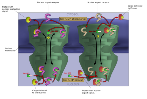
The movement of macromolecules across the nuclear envelope is an active process known as the Ran-GTP nuclear transport cycle. This system uses specific transport receptors to shuttle proteins and RNA in and out of the nucleus based on signal sequences.
Replication and Gene Expression
The nucleus is the site of DNA replication and the first stage of gene expression: transcription. Here, DNA is transcribed into pre-mRNA, which is then processed (spliced) before being exported to the cytoplasm for translation into proteins.
Nuclear Variation and Evolution
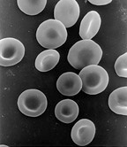
While most animal cells contain a single nucleus, there are notable exceptions. Some cells are anucleated (lacking a nucleus), such as mature mammalian red blood cells, which expel their nuclei during development to maximize space for hemoglobin.
Conversely, some cells are multinucleated. From an evolutionary perspective, the origin of the nucleus remains a subject of scientific debate, with theories ranging from the internal folding of the plasma membrane to viral ancestry.
Summary of Nuclear Bodies
| Structure Name | Approximate Diameter |
|---|---|
| Cajal bodies | 0.2–2.0 μm |
| Clastosomes | 0.2–0.5 μm |
| PML bodies | 0.2–1.0 μm |
| Paraspeckles | 0.5–1.0 μm |
| PIKA | 5 μm |
| Speckles | 20–25 nm |
Frequently Asked Questions
What is the difference between chromatin and chromosomes?
Chromatin is the relaxed form of DNA and proteins found in the nucleus during interphase. Chromosomes are the highly condensed versions of chromatin that become visible during cell division (mitosis).
How do molecules enter and exit the nucleus?
Molecules move through nuclear pores via the Ran-GTP nuclear transport cycle, which uses specialized transport proteins to actively move macromolecules across the nuclear envelope.
Why do red blood cells lack a nucleus?
In mammals, red blood cells expel their nuclei during development to create more room for hemoglobin, which allows the cell to transport oxygen more efficiently.
What is the role of the nucleolus?
The nucleolus is responsible for the synthesis of ribosomal RNA (rRNA) and the assembly of ribosome subunits, which are later exported to the cytoplasm to synthesize proteins.
What is the function of the nuclear lamina?
The nuclear lamina provides structural rigidity to the nucleus, helps organize the positioning of chromatin, and is involved in the regulation of DNA replication.