bird anatomyavian skeletal systempneumatized bonesavian respiratory systempectoralis major

Bird Anatomy: Biological Adaptations for Flight and Survival

Bird Anatomy: Biological Adaptations for Flight and Survival Bird anatomy is a masterclass in evolutionary engineering, characterized by a suite of physiological structures specifically a...

Bird Anatomy: Biological Adaptations for Flight and Survival

Bird anatomy is a masterclass in evolutionary engineering, characterized by a suite of physiological structures specifically adapted for the demands of flight. To achieve lift and sustain high-energy movement, birds have evolved a lightweight skeletal system, powerful musculature, and highly efficient circulatory and respiratory systems capable of supporting extreme metabolic rates.

Beyond the mechanics of flight, the development of the beak and specialized internal organs allows birds to occupy diverse ecological niches across the globe. From the acute vision of raptors to the unique air sac system that ensures a constant flow of oxygen, every aspect of avian biology is tuned for efficiency.
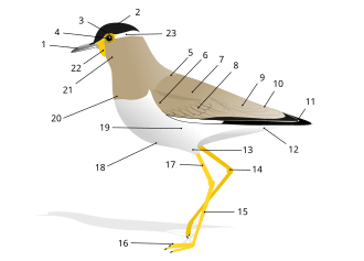
External anatomy (topography) of a typical bird (in this case, a yellow wattled lapwing):BeakHeadIrisPupilMantleLesser covertsScapularsCovertsTertialsRumpPrimariesVentThighTibio-tarsal articulationTarsusFeetTibiaBellyFlanksBreastThroatWattleEyestripe

Key Facts

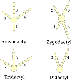
Four types of bird feet
  • Pneumatized Bones: Many bird bones are hollow and reinforced with internal struts to reduce weight without sacrificing strength.
  • Flight Muscles: The pectoralis major and supracoracoideus can account for 25–40% of a bird's total body weight.
  • Respiratory Efficiency: Avian lungs are supplemented by air sacs, providing a respiratory surface area approximately 15% greater than that of similar-sized mammals.
  • Acute Vision: Some raptors possess photoreceptor densities up to 1,000,000 per square mm, making their vision eight times sharper than humans.
  • Unique Immune Organ: The bursa of Fabricius is a specialized pouch connected to the cloaca that houses B lymphocytes.

The Skeletal System
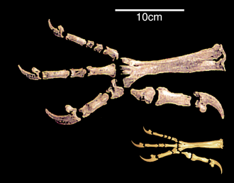
Comparative morphology of the paw skeleton of the extinct Haast's eagle with its closest living relative the little eagle.

The avian skeleton is designed for maximum lightness and structural integrity. Many bones are pneumatized, meaning they are hollow and contain criss-crossing struts for strength. These hollow spaces often house respiratory air sacs. While large soaring birds typically have the most pneumatized bones, diving birds (such as penguins, loons, and puffins) and kiwis lack them entirely to assist with buoyancy and diving.

The Axial Skeleton

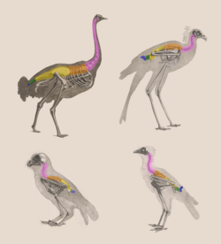
The vertebral column of a bird is divided into five distinct sections: the cervical vertebrae (neck), thoracic vertebrae, the synsacrum (a fused section of the lower spine), caudal vertebrae, and the pygostyle (the final fused vertebrae supporting tail feathers).
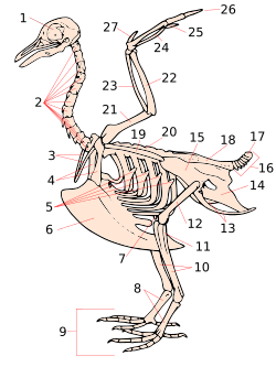
A stylised dove skeleton. skullcervical vertebraefurculacoracoiduncinate processes of ribskeelpatellatarsometatarsusdigitstibia (tibiotarsus)fibula (tibiotarsus)femurischium (innominate)pubis (innominate)ilium (innominate)caudal vertebraepygostylesynsacrumscapuladorsal vertebraehumerusulnaradiusCarpometacarpusDigit IIIDigit IIDigit I (alula)
Collage of bird anatomical illustrations with the different vertebral sections color-coded across various species. The species included are as follows: Top row (left to right) Struthio camelus and Sagittarius serpentarius (formerly Gypogeranus serpentarius) Bottom row (left to right) Megascops choliba decussatus (formerly known as Strix decussata) and Falco rusticolus islandus (formerly Falco islandus). Sections of the vertebral column in anatomical bird diagrams Color Vertebral section Pink Cervical vertebrae Orange Thoracic/dorsal vertebrae Yellow Synsacrum Green Caudal vertebrae Blue Pygostyle

Pelvic Girdle and Skull

The pelvic girdle integrates the lower vertebral column to provide a stable base for landing and locomotion. The skull is characterized by the loss of teeth and the evolution of the beak, a lightweight yet strong structure that serves as a primary tool for feeding and manipulation.
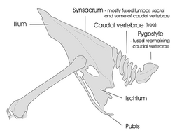

Diagram of a general bird pelvic girdle skeleton including the lower vertebral column sections. The diagram includes the synsacrum, caudal, and pygostyle vertebrae. Note that the caudal vertebrae (5–10) are not fused in this diagram but can be in certain species.
Diagram of a general bird pelvic girdle skeleton including the lower vertebral column sections. Note that the caudal vertebrae (5–10) are not fused in this diagram but can be in certain species.
The typical cranial anatomy of a bird. Pmx= premaxilla, M= maxilla, D= dentary, V= vomer, Pal= palatine, Pt= Pterygoid, Lc= Lacrimal

Muscular System and Flight Mechanics
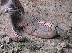
Ostrich foot integument (podotheca)

Most birds possess roughly 175 muscles, with the majority of muscle mass concentrated on the ventral (belly) side to maintain a low center of gravity. The most critical muscles for flight are the pectorals (pectoralis major), which provide the powerful downstroke and comprise 15–25% of the body weight.

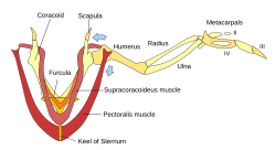
Deep to the pectorals lies the supracoracoideus (pectoralis minor), which acts as a pulley system to lift the wing. Both of these muscle groups attach to the keel, an enlarged extension of the sternum (breastbone) that provides the necessary surface area for such massive muscle attachments.
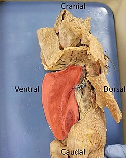
Highlighted in red is an intact keeled sternum of a dissected pigeon. In flying birds the sternum is enlarged for increased muscle attachment.
The supracoracoideus works using a pulley-like system to lift the wing while the pectorals provide the powerful downstroke
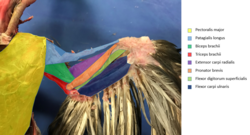
Labelled ventral musculature of a pigeon wing
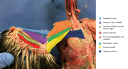
Labelled dorsal musculature of a pigeon wing

Respiratory and Circulatory Systems
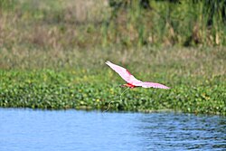
A Roseate spoonbill excreting urine in flight

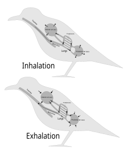
Birds possess one of the most efficient respiratory systems in the animal kingdom. While their lungs are smaller than those of mammals, they are supported by a complex system of air sacs that occupy about 15% of the total body volume. This system allows for a unidirectional flow of air, ensuring that oxygen-rich air passes through the lungs during both inhalation and exhalation.
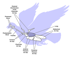
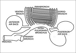
The arrangement of the air sacs and lungs in birds
The anatomy of bird's respiratory system, showing the relationships of the trachea, primary and intra-pulmonary bronchi, the dorso- and ventro-bronchi, with the parabronchi running between the two. The posterior and anterior air sacs are also indicated, but not to scale.
Inhalation–exhalation cycle in birds.
The cross-current respiratory gas exchanger in the lungs of birds. Air is forced from the air sacs unidirectionally (from right to left in the diagram) through the parabronchi. The pulmonary capillaries surround the parabronchi in the manner shown (blood flowing from below the parabronchus to above it in the diagram).[59][61] Blood or air with a high oxygen content is shown in red; oxygen-poor air or blood is shown in various shades of purple-blue.

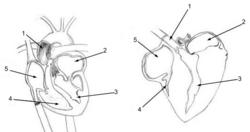
To support this high oxygen demand, the avian heart pumps faster than the mammalian heart. Consequently, the muscles surrounding the ventricles are thicker to handle the increased pressure. In birds, the superior cava is double, further enhancing blood return to the heart.

The human heart (left) and chicken heart (right) share many similar characteristics. Avian hearts pump faster than mammalian hearts. Due to the faster heart rate, the muscles surrounding the ventricles of the chicken heart are thicker. Both hearts are labeled with the following parts: 1. Ascending Aorta 2. Left Atrium 3. Left Ventricle 4. Right Ventricle 5. Right Atrium. In chickens and others birds, the superior cava is double.

Digestive and Urogenital Systems

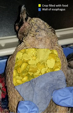
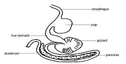
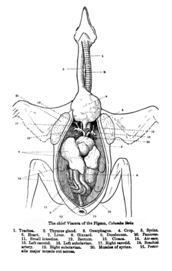
The avian digestive tract is adapted for rapid processing. A key feature is the crop, an out-pouching of the esophagus used to store food before digestion. Food then moves through the proventriculus (glandular stomach) to the gizzard, where it is mechanically ground.

Pigeon crop containing ingested food particles is highlighted in yellow. The crop is an out-pouching of the esophagus and the wall of the esophagus is shown in blue.
Simplified depiction of avian digestive system.
Alimentary canal of the bird exposed

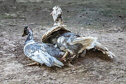
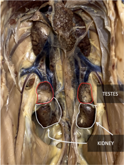
The urogenital system includes kidneys located along the medial spine. In females, the reproductive system typically involves a single functional oviduct leading to the cloaca. In some species, such as ducks, males possess a corkscrew-shaped penis for copulation.

Diagram of a female chicken reproductive system A. Mature ovum, B. Infundibulum, C. Magnum, D. Isthmus, E. Uterus, F. Vagina, G. Cloaca, H. Large intestine, I. rudiment of right oviduct
Copulating ducks showing a corkscrew penis inserted in a cloaca
Chicken's kidneys visualized at the bottom of the abdomen cavity, along the medial spine of the chicken. Testes are labeled above the kidneys.

Sensory Organs and the Nervous System

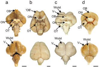
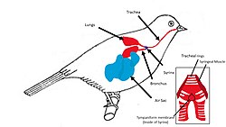
Birds are renowned for their acute eyesight. This is achieved through high densities of photoreceptors in the retina and a high number of neurons in the optic nerves. Some species have an indented fovea that magnifies the central visual field, and others, like hummingbirds, possess two foveas per eye. Additionally, birds produce sound via the syrinx, a specialized vocal organ located at the base of the trachea.

Brains of an emu, a kiwi, a barn owl, and a pigeon, with visual processing areas labelled
Vocal Bird anatomy: Birds produce sounds through the air that passes through the Syrinx, which is shown close up in the bottom right.

Immune System: The Bursa of Fabricius

A unique feature of avian anatomy is the bursa of Fabricius, a circular pouch connected to the cloaca. This organ is essential for the development of the immune system, specifically housing B lymphocytes within follicles consisting of a cortex and a medulla. These lymphocytes are critical for the bird's ability to produce antibodies.

Summary of Avian Anatomical Adaptations
System Key Feature Primary Function
Skeletal Pneumatized Bones Weight reduction for flight
Muscular Keeled Sternum Attachment for powerful flight muscles
Respiratory Air Sacs Unidirectional, continuous oxygen flow
Digestive Crop & Gizzard Food storage and mechanical grinding
Sensory High Photoreceptor Density Extreme visual acuity (especially in raptors)

Frequently Asked Questions

Why are bird bones hollow?

Many bird bones are pneumatized (hollow) to reduce the overall body weight, making it easier to achieve and maintain flight, while internal struts provide the necessary structural strength.

How do birds breathe differently than mammals?

Unlike mammals, birds use a system of air sacs that act as bellows to push air unidirectionally through the lungs. This ensures that the lungs receive a constant supply of fresh, oxygenated air during both inhalation and exhalation.
What is the purpose of the keel on a bird's sternum?

The keel is an enlarged ridge on the breastbone that provides a large surface area for the attachment of the pectoralis major and supracoracoideus muscles, which are responsible for the wing's downstroke and upstroke.

What is the bursa of Fabricius?

The bursa of Fabricius is a specialized immune organ in birds, located near the cloaca, where B lymphocytes mature and develop, enabling the bird to fight infections.

How is bird vision superior to human vision?

Birds, particularly raptors, have a much higher density of photoreceptors in their retinas and more neurons in their optic nerves. Some also have specialized foveas that magnify their central field of vision.