Abdominal Aortic Aneurysm
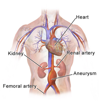
An abdominal aortic aneurysm (AAA) occurs when a section of the abdominal aorta—the body's largest artery—becomes weakened and bulges outward. This condition is particularly dangerous because it often develops without symptoms until the aneurysm grows large enough to risk a rupture, which can be fatal. Managing AAA requires a combination of vigilant screening, risk factor modification, and timely surgical intervention.
Medical professionals in vascular surgery and interventional radiology typically manage these cases, focusing on monitoring the diameter of the aorta to determine when the risk of rupture outweighs the risks of surgical repair.
Key Facts
- Definition: An abdominal aorta diameter greater than 3 cm is diagnostic of an aneurysm.
- Primary Demographic: Most common in males over the age of 50.
- Prevalence: Affects approximately 5% of males over 65 years old.
- Major Risk Factors: Smoking, hypertension, cardiovascular disease, family history, and Marfan syndrome.
- Critical Danger: A rupture can lead to massive internal bleeding; 168,200 deaths were attributed to aortic aneurysms in 2015.
Symptoms and Diagnosis
One of the most challenging aspects of an AAA is that it is frequently asymptomatic. Many patients have no warning signs until the aneurysm ruptures. However, some individuals may experience pain in the abdomen, back, or legs.
Diagnosis is primarily achieved through medical imaging. Ultrasonography (ultrasound) is often used for screening and monitoring, while CT scans provide detailed measurements and help surgeons plan interventions. A diagnosis is confirmed when the abdominal aorta diameter is measured at > 3 cm.
Aneurysm Classification by Size
Clinicians classify the severity of an AAA based on its diameter to determine the appropriate management strategy:
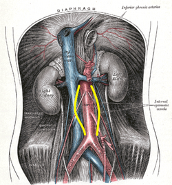
- Ectatic or Mild Dilatation: Between 2.0 cm and 3.0 cm.
- Moderate: Between 3.0 cm and 5.0 cm.
- Large or Severe: Greater than 5.0 cm or 5.5 cm.
Risk Assessment and Rupture Probability
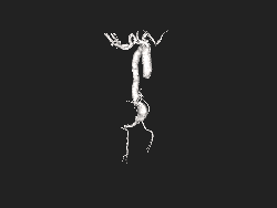
The risk of a rupture increases significantly as the aneurysm grows. While diameter is the primary metric, the growth rate per year also plays a critical role in determining the urgency of surgery.
| AAA Size (cm) | Average Growth Rate (cm/yr) | Annual Rupture Risk (%) |
|---|---|---|
| 3.0–3.9 | 0.39 | 0% |
| 4.0–4.9 | 0.36 | 0.5–5% |
| 5.0–5.9 | 0.43 | 3–15% |
| 6.0–6.9 | 0.64 | 10–20% |
| ≥7.0 | - | 20–50% |
Prevention and Management
Preventing the development or progression of an AAA focuses on controlling cardiovascular health. The most effective measures include smoking cessation and the aggressive treatment of hypertension (high blood pressure).
Screening Guidelines
Screening intervals vary by organization (such as SVS, ESVS, and RESCAN) and depend on the initial size of the aneurysm. For example, for an aneurysm sized 3.0–3.9 cm, screening may occur every 3 years for men and every 2 to 3 years for women. As the size increases toward 5.0 cm, the interval shortens to 6 months or leads directly to surgical consideration.
Treatment Options
When an aneurysm reaches a critical size or grows rapidly, surgery is required. There are two primary methods:
- Open Repair: A traditional surgical procedure where the weakened section of the aorta is replaced with a synthetic graft.
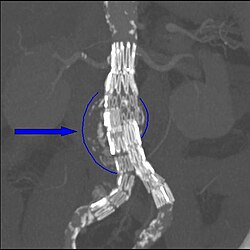
- Endovascular Aneurysm Repair (EVAR): A less invasive procedure where a stent-graft is inserted through the arteries (usually in the groin) and deployed inside the aorta to reinforce the wall.
Frequently Asked Questions
What is the main cause of an abdominal aortic aneurysm?
While multiple factors contribute, the primary risk factors include smoking, hypertension, other cardiovascular diseases, a family history of the condition, and genetic disorders such as Marfan syndrome.
How is an AAA diagnosed?
Diagnosis is made using medical imaging, most commonly abdominal ultrasonography or CT scans. An aneurysm is clinically defined as an abdominal aorta diameter exceeding 3 cm.
Can an AAA be treated without surgery?
Small aneurysms are often managed conservatively through regular monitoring (surveillance) and the treatment of risk factors like high blood pressure and smoking. However, once an aneurysm reaches a certain size (typically ≥5.0–5.5 cm), surgery is generally recommended to prevent rupture.
What is the difference between open repair and endovascular repair?
Open repair involves a major surgical incision to manually replace the damaged aorta with a graft. Endovascular repair (EVAR) is a minimally invasive approach that uses a catheter to place a stent-graft inside the artery, reducing recovery time and surgical trauma.
Who should be screened for AAA?
Screening is particularly important for males over the age of 65, as they have a higher prevalence of the condition (approximately 5%). Individuals with a family history or those who have smoked are also high-priority candidates.