Visual Field: Anatomy, Measurement, and Clinical Defects
The visual field is the entire area that a person can see when their eyes are fixed in one direction. While often used interchangeably with the "field of view" (FOV) in optical instruments, in medical contexts, it refers specifically to the functional capacity of vision recorded through specialized testing. For psychologists, it is viewed as a perceptual array of sensations available during introspection.
Understanding the boundaries and structure of the visual field is critical for diagnosing diseases of the eye, optic nerve, and brain. By mapping where vision is clear and where it is diminished, clinicians can pinpoint the exact location of neurological or ocular damage.
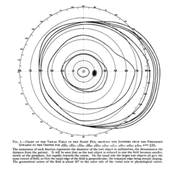
Normal Limits of Human Vision
Human vision is divided into monocular (single eye) and binocular (both eyes) fields. A single eye's visual field typically extends:
- Nasally (toward the nose): Approximately 60 degrees.
- Temporally (away from the nose): Approximately 107 degrees.
- Superiorly (above): Approximately 70 degrees.
- Inferiorly (below): Approximately 80 degrees.
The binocular visual field is the combined area of both eyes. This overlap allows for depth perception and a wider horizontal reach. Interestingly, while the nose occupies part of this field, the brain processes this information to ensure the nose is not consciously noticed during daily tasks.
Central Vision Zones
The center of the visual field is responsible for high-resolution detail. This is divided into three primary zones:
- Macula: The central 17 degrees of the field.
- Fovea: The central 5.2 degrees.
- Foveola: The innermost 1–1.2 degrees (sometimes referred to as the "clinical fovea").
Key Facts
- Binocular Field: The superimposition of both monocular fields; EU driving requirements typically mandate a total horizontal field of 120 degrees.
- Perimetry: The primary clinical method used to measure the visual field.
- Scotoma: A localized area of partial or complete vision loss within the visual field.
- Tunnel Vision: A severe loss of peripheral vision with the retention of central vision.
- Hemianopia: Loss of half of the visual field, often indicating a lesion after the optic chiasm.
Measuring the Visual Field
Clinicians use perimetry to map the visual field. This can be kinetic (moving a light spot until the patient sees it) or static (flashing lights of varying intensity at fixed points). Common devices include the Humphrey Field Analyzer, Octopus, and Heidelberg Edge Perimeter. For central field measurements, a campimeter is often used.
A simpler, rapid assessment is confrontational field testing. In this method, a practitioner holds up fingers in different quadrants of the patient's vision. If the patient reports them accurately, the result is recorded as "full to finger counting" (FTFC). This method can also identify the blind spot by observing when a small object disappears from view.
Visual Field Defects and Their Causes
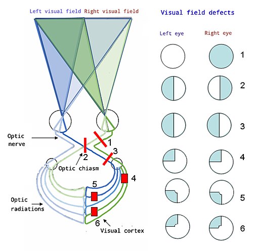
Vision loss is categorized by the location of the lesion. Defects can be heteronymous (affecting different sides of the two eyes) or homonymous (affecting the same side of both eyes).
| Lesion Location | Typical Defect | Example Condition |
|---|---|---|
| Retina / Eye | Central or Peripheral Scotoma | Glaucoma, AMD |
| Optic Nerve | Heteronymous defects / Total loss in one eye | Optic Neuritis |
| Optic Chiasm | Bitemporal Hemianopia | Pituitary Tumor |
| Post-Chiasm (Tract/Radiation) | Homonymous Hemianopia / Quadrantanopia | Stroke, Brain Injury |
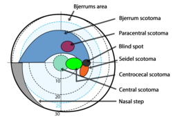
Glaucoma and the Retinal Nerve Fiber Layer
In glaucoma, damage to the retinal nerve fiber layer (RNFL) creates distinct patterns. Early changes often appear in Bjerrum's area (10°–20° from fixation). These progress from small wing-shaped scotomas to Siedel's sickle-shaped scotoma, and eventually to arcuate scotomas. In advanced stages, these may merge into a ring or double arcuate scotoma, or result in tubular vision (tunnel vision), where only the central macular fibers remain functional.
Macular Degeneration (AMD)
Age-related Macular Degeneration affects the macula, the area responsible for high-resolution vision and reading. This typically results in central scotomas or metamorphopsia (distorted vision), leaving the peripheral field intact.
Neurological and Other Causes
- Cerebral Palsy: May cause hemianopia, central/peripheral scotomas, or islands of vision.
- Periventricular Leukomalacia (PVL): Often results in bilateral inferior visual field defects.
- Cataracts: Generally cause a generalized depression of the entire field.
- Retinitis Pigmentosa: A primary cause of tubular vision.
Frequently Asked Questions
What is the difference between a scotoma and hemianopia?
A scotoma is a localized area of vision loss (a "blind spot") within the visual field, whereas hemianopia is the loss of an entire half of the visual field in one or both eyes.
What is bitemporal hemianopia?
Bitemporal hemianopia is the loss of the outer (temporal) half of the visual field in both eyes, typically caused by a lesion in the center of the optic chiasm.
How is "tunnel vision" different from a central scotoma?
Tunnel vision (tubular vision) is the loss of peripheral vision with the center remaining clear. A central scotoma is the opposite: the center is blind, but the peripheral vision remains.
What is the "blind spot" in the visual field?
The blind spot is a natural area of the visual field where the optic nerve exits the eye, meaning there are no photoreceptors present to detect light.
What does "full to finger counting" mean in a medical report?
This is a result from a confrontational field test indicating that the patient can see the practitioner's fingers in all quadrants, suggesting a generally normal visual field.