Uric Acid: Chemistry, Biological Function, and Clinical Significance
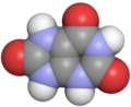
Uric acid (IUPAC name: 7,9-Dihydro-1H-purine-2,6,8(3H)-trione) is a critical metabolic byproduct of purine degradation in humans. While it serves as a natural component of the body's biochemistry, its concentration in the blood and urine is a vital indicator of renal health and metabolic balance. When these levels deviate from the norm, it can lead to a variety of clinical conditions, most notably gout.
In the human body, uric acid is the final product of a metabolic pathway that breaks down purines. The sequence follows a specific chemical progression: AMP → IMP → Inosine → Hypoxanthine → Xanthine → Uric Acid.

Chemical Properties and Structure
Uric acid appears as white crystals with a molar mass of 168.112 g·mol. It is characterized by its low solubility in water (6 mg/100 mL at 20 °C) and a melting point of 300 °C. Chemically, it exists in several forms, including the lactam (a stable tautomer), the lactim, and the urate ion, which is its conjugate base.

The acidity of uric acid is defined by a pKa of 5.6, while its basicity is marked by a pKb of 8.4. Because of its chemical nature, it often forms various salts, known as urates, which vary significantly in their solubility depending on the cation involved.

Solubility of Urate Salts
The solubility of uric acid is highly dependent on the salt form. For instance, sodium hydrogen urate is significantly more soluble in cold water than disodium urate, which affects how these substances precipitate in human tissues.
| Compound | Cold Water | Boiling Water |
|---|---|---|
| Uric acid | 15,000 | 2,000 |
| Sodium hydrogen urate | 1,175 | 124 |
| Potassium hydrogen urate | 790 | 75 |
| Calcium urate | 1,500 | 1,440 |
| Barium urate | 7,900 | 2,700 |
Physiology and Genetic Influence
In humans, the kidneys are responsible for approximately 70% of daily uric acid disposal. Normal urinary excretion ranges from 270 to 360 mg per day. In the blood plasma, reference ranges typically vary by sex: men generally range from 3.4 to 7.2 mg per 100 mL, while women range from 2.4 to 6.1 mg per 100 mL.
While diet—specifically meat and seafood—can influence serum urate levels, genetic variation is a more significant contributor. Mutations in urate transport proteins can impair renal excretion, leading to high blood levels. Key genes linked to serum urate include SLC2A9 (which encodes GLUT9, a transporter for both uric acid and fructose), ABCG2, SLC17A1, and SLC22A12, among others.
Clinical Significance
Deviations in uric acid levels are categorized based on whether they occur in the blood (serum) or urine.
- Hyperuricemia: Excess uric acid in the blood.
- Hypouricemia: Abnormally low uric acid in the blood.
- Hyperuricosuria: Excess uric acid in the urine.
- Hypouricosuria: Abnormally low uric acid in the urine.
Gout and Crystal Formation
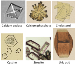
Hyperuricemia can lead to gout, a painful inflammatory condition. This occurs when monosodium urate crystals precipitate as needle-like structures in joints, capillaries, and skin. While gout often occurs at serum levels above 6 mg per 100 mL, some individuals may remain asymptomatic even with levels as high as 9.6 mg per 100 mL.
Other Related Conditions
Beyond gout, abnormal uric acid levels are associated with several other medical conditions:
- Tumor Lysis Syndrome: Rapid breakdown of cancer cells releasing high levels of purines.
- Lesch-Nyhan Syndrome: A genetic disorder causing overproduction of uric acid.
- Renal Health: Formation of uric acid stones in the urinary tract.
- Systemic Links: Potential associations with cardiovascular disease and diabetes.
Key Facts
- Chemical Formula: C5H4N4O3.
- Primary Excretion: 70% of uric acid is removed via the kidneys.
- Gout Trigger: Caused by the precipitation of monosodium urate crystals in tissues.
- Genetic Drivers: Genes like SLC2A9 play a larger role in serum levels than diet alone.
- Normal Range: Typically 3.4–7.2 mg/100 mL for men and 2.4–6.1 mg/100 mL for women.
Frequently Asked Questions
What is the difference between hyperuricemia and gout?
Hyperuricemia is the medical term for having high levels of uric acid in the blood. Gout is the clinical condition that results when those high levels cause urate crystals to precipitate in the joints and tissues, leading to pain and inflammation.
Does diet cause high uric acid levels?
While purine-rich foods like seafood and meat can elevate levels, genetic mutations in transport proteins (such as those encoded by the SLC2A9 gene) are often more significant contributors to chronic hyperuricemia.
How is uric acid excreted from the body?
The majority (about 70%) of uric acid is excreted through the kidneys into the urine. A smaller portion is eliminated through the intestinal tract.
What are the normal blood levels of uric acid?
For men, the typical reference range is 3.4 to 7.2 mg per 100 mL. For women, the range is generally 2.4 to 6.1 mg per 100 mL.
What is the role of the SLC2A9 gene?
The SLC2A9 gene encodes the GLUT9 transporter, which is responsible for transporting both uric acid and fructose, significantly influencing how the kidneys excrete urate.