Tongue Anatomy: Structure, Function, and Biological Complexity
The human tongue is a sophisticated muscular organ essential to several vital biological processes, including speech, mastication (chewing), and gustation (the sense of taste). As a key component of both the alimentary tract and the gustatory system, it serves as a bridge between the oral cavity and the pharynx, facilitating the movement of food and the articulation of complex sounds.
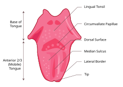
Anatomically, the tongue is divided into two primary regions: the oral part at the front and the pharyngeal part at the back. A vertical section of fibrous tissue known as the lingual septum separates the left and right sides, creating a visible groove on the surface called the median sulcus.
Key Facts
- Average Length: Approximately 10 cm from the tip to the oropharynx.
- Average Weight: 99g in adult males and 79g in adult females.
- Muscle Groups: Comprised of four intrinsic muscles (shape) and four extrinsic muscles (position).
- Primary Blood Supply: Provided mainly by the lingual artery.
- Sensory Division: The anterior two-thirds and posterior one-third have distinct nerve supplies for taste and sensation.
Detailed Anatomical Structure
The Upper and Lower Surfaces
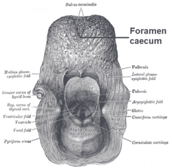
The upper surface, or dorsum, is characterized by various projections called papillae. These papillae include filiform, fungiform, vallate, and foliate types. Notably, only the filiform papillae lack taste buds. The dorsum is divided by the median sulcus, which ends at the foramen cecum. This point marks the beginning of the terminal sulcus, a V-shaped groove that separates the anterior oral part from the posterior pharyngeal part.
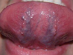
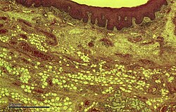
The undersurface, or ventral surface, is a smooth, non-keratinized epithelium. It features the frenulum, a fold of mucous membrane that tethers the tongue to the floor of the mouth. On either side of the frenulum are the sublingual caruncles, which serve as drainage points for the submandibular salivary glands.
Muscular Composition
The tongue's movement is driven by eight distinct muscles, categorized by their attachment points:
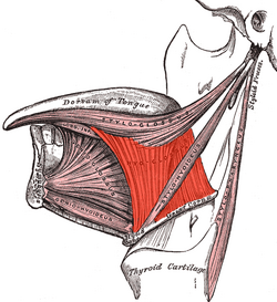
Extrinsic Muscles (Positioning)These muscles are anchored to bone and are responsible for moving the tongue within the mouth:
- Genioglossus: Arises from the mandible; known as the "safety muscle" because it protrudes the tongue forward.
- Hyoglossus: Arises from the hyoid bone; functions to retract and depress the tongue.
- Styloglossus: Arises from the styloid process; draws the sides of the tongue up to create a swallowing trough.
- Palatoglossus: Arises from the palatine aponeurosis; elevates the back of the tongue during swallowing.
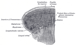
These muscles originate and insert within the tongue itself, allowing it to change shape without bone attachment:
- Superior longitudinal muscle: Shortens and curls the tongue upward.
- Inferior longitudinal muscle: Shortens and curls the tongue downward.
- Vertical muscle: Flattens the tongue.
- Transverse muscle: Lengthens and narrows the tongue.
Nerve Supply and Sensory Perception
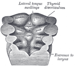
The innervation of the tongue is complex due to its embryological origins from different pharyngeal arches. This results in a split sensory map:
- Anterior Two-Thirds: Taste is managed by the chorda tympani (a branch of the facial nerve), while general sensation is handled by the lingual nerve (a branch of the trigeminal nerve).
- Posterior One-Third: Both taste and sensation are supplied by the glossopharyngeal nerve.
- Base of Tongue: Taste and sensation are managed by the internal branch of the superior laryngeal nerve (a branch of the vagus nerve).
Motor control for nearly all muscles is provided by the hypoglossal nerve (CN XII), with the exception of the palatoglossus, which is controlled by the vagus nerve.
Circulation and Lymphatic Drainage
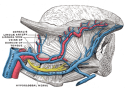
The primary blood supply is the lingual artery, a branch of the external carotid artery. Secondary supply to the root comes from the tonsillar branch of the facial artery and the ascending pharyngeal artery. For lymphatic drainage, the tip of the tongue drains to the submental nodes, the anterior two-thirds to the submandibular nodes, and the posterior third to the jugulo-omohyoid nodes.
Summary of Tongue Anatomy
| Feature | Description/Function | Key Component |
|---|---|---|
| Muscles (Shape) | Intrinsic muscles located within the organ | Superior/Inferior longitudinal, Vertical, Transverse |
| Muscles (Position) | Extrinsic muscles anchored to bone | Genioglossus, Hyoglossus, Styloglossus, Palatoglossus |
| Sensory (Anterior) | Taste and general sensation (front 2/3) | Facial nerve (taste), Trigeminal nerve (sensation) |
| Sensory (Posterior) | Taste and general sensation (back 1/3) | Glossopharyngeal nerve |
| Blood Supply | Primary arterial source | Lingual artery |
Frequently Asked Questions
What is the difference between apical and laminal sounds?
In phonetics, apical sounds are produced using the tip of the tongue, whereas laminal sounds are produced using the blade (the area just behind the tip).
What is ankyloglossia?
Ankyloglossia, commonly known as tongue-tie, is a congenital disorder where a short, thickened frenulum tethers the tongue to the floor of the mouth, potentially affecting speech, eating, and swallowing.
How does the tongue develop in an embryo?
Development begins in the fourth week from the tuberculum impar (median tongue bud). By the fifth week, lateral lingual swellings expand to cover this bud, forming the anterior two-thirds of the tongue.
Why do different animals have different tongue structures?
Tongue structures vary significantly across species to suit specific biological needs, such as the specialized tongue of a giraffe or the extended proboscis of a Macroglossum moth.