SPDM: Advancing Biological Imaging through Spectral Precision Distance Microscopy
In the world of microscopy, the ability to see the smallest structures of life is often limited by the laws of physics. Conventional optical resolution is typically capped at approximately 200-250 nm, meaning any two objects closer than this distance appear as a single, blurry spot. However, Spectral Precision Distance Microscopy (SPDM) is a family of localization microscopy techniques designed to break this barrier, allowing scientists to measure distances with nanometer accuracy.
The Challenge of Optical Resolution
When a single, tiny source of light is viewed through a microscope, it does not appear as a perfect point. Instead, it creates a blurry spot known as the point spread function (PSF). While this blurriness seems like a limitation, computer algorithms can analyze the PSF and the noise properties of the detector to calculate the exact center of that spot with high precision.
The problem arises when multiple light sources are crowded together. If too many sources are within the same resolution limit, their blurry spots overlap and merge, making it impossible for algorithms to distinguish one from another.
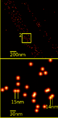
How SPDM Works
SPDM overcomes the problem of overlapping signals by ensuring that only a few sources are measured at any given time. By making each source optically isolated—meaning they are separated by more than the standard 200-250 nm resolution limit—the system can accurately locate the center of each individual spot.
Achieving Optical Isolation
To isolate these sources, SPDM leverages the unique characteristics of fluorescent molecules. This is achieved through several methods:
- Spectral Variation: Using specific light sources and filters to target molecules with different absorption or emission spectra.
- Fluorescent Lifetime: Distinguishing molecules based on the duration they remain in an excited state.
- Advanced Filtering: Utilizing other subtle molecular signatures to separate signals.
Topological Resolution and Precision
The effectiveness of SPDM is measured by its topological resolution, which is the smallest measurable distance between two particles with different spectral characteristics. While the physical point spread function remains the same, the ability to localize the center of these particles allows for an effective optical resolution several times better than conventional limits.
This breakthrough is rooted in foundational work from 1996, which provided the first experimental and theoretical proof that fluorescent objects could be localized in 3D space with an accuracy of approximately 1 nm. This represents a precision better than 1/100 of the wavelength of visible light.
Key Facts
- Conventional Limit: Standard optical resolution is approximately 200-250 nm.
- Localization Accuracy: SPDM can achieve precision in the range of 1 nm.
- Core Mechanism: It uses spectral signatures to optically isolate light sources.
- Primary Applications: Essential for genome research (functional organization) and membrane structure analysis.
- Technical Basis: Relies on calculating the center of the point spread function (PSF).
Comparison of Microscopy Resolutions
| Feature | Conventional Microscopy | SPDM (Localization Microscopy) |
|---|---|---|
| Typical Resolution Limit | 200-250 nm | Nanometer scale (down to ~1 nm) |
| Image Appearance | Blurry spots (PSF) | Calculated precise coordinates |
| Handling Dense Sources | Sources blur together | Sources are optically isolated |
| Requirement for Precision | Standard optics | Spectral signatures & laser precision |
Frequently Asked Questions
What is the point spread function (PSF)?
The point spread function is the blurry spot produced by a microscope when imaging a single, infinitesimal point source of light. SPDM uses the PSF as a baseline to calculate the exact center of a molecule's position.
How does SPDM differ from standard fluorescence microscopy?
Standard microscopy is limited by the diffraction of light (approx. 200-250 nm). SPDM bypasses this by isolating individual molecules spectrally and using mathematical algorithms to locate them with nanometer precision.
What are the main applications of SPDM?
SPDM is primarily used in high-level biological research, specifically in studying the functional organization of the genome and investigating the intricate structures of cellular membranes.
What is topological resolution?
Topological resolution refers to the smallest measurable distance between two particles that have different spectral characteristics, effectively providing a much higher resolution than classical optical limits.
Can SPDM locate objects in 3D?
Yes, the foundations of localization microscopy established in 1996 proved that fluorescent objects could be localized in the nanoscale across three dimensions.