Proteases: The Biological Engines of Protein Degradation
Proteases, also known as peptidases or proteinases, are specialized enzymes that drive the process of proteolysis. This critical biological function involves breaking down proteins into smaller polypeptides or individual amino acids, which can then be used to create new protein products. By cleaving the peptide bonds that hold amino acids together through a process called hydrolysis—a chemical reaction where water is used to break bonds—proteases enable essential life processes.
Without these biological accelerators, the natural breakdown of proteins would be impossibly slow, potentially taking hundreds of years. Because they are so vital, proteases have evolved independently multiple times across all forms of life, from simple viruses to complex eukaryotes. They are central to digestion, the recycling of old proteins (protein catabolism), and intricate cell signaling pathways.
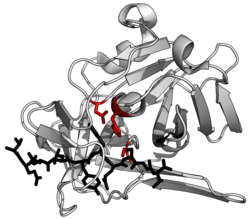
Key Facts
- Function: Catalyze the cleavage of peptide bonds via hydrolysis or elimination reactions.
- Ubiquity: Found in all living organisms, including viruses, bacteria, archaea, plants, and animals.
- Diversity: Classified into seven broad groups based on their catalytic residues.
- Regulation: Controlled by protease inhibitors and through autolysis (self-cleavage).
- Medical Impact: Involved in blood clotting, apoptosis, and cancer metastasis.
Classification of Proteases
Proteases are categorized using several different systems depending on whether the focus is on their chemical mechanism, their evolutionary history, or their environmental preferences.
Based on Catalytic Residues
The most common classification is based on the specific amino acid or cofactor used to perform the cleavage. Most proteases make a residue or a water molecule nucleophilic (capable of attacking an electron-deficient center) to break the peptide carbonyl group.
- Serine proteases: Use a serine alcohol.
- Cysteine proteases: Use a cysteine thiol.
- Threonine proteases: Use a threonine secondary alcohol.
- Aspartic proteases: Use an aspartate carboxylic acid.
- Glutamic proteases: Use a glutamate carboxylic acid.
- Metalloproteases: Use a metal ion, typically zinc.
- Asparagine peptide lyases: Use asparagine to perform an elimination reaction that does not require water.
Many of these enzymes utilize a catalytic triad, where a histidine residue activates the serine, cysteine, or threonine to act as the nucleophile. In contrast, metalloproteases, aspartic, and glutamic proteases activate a water molecule to attack the bond.
Evolutionary Phylogeny
The MEROPS database provides a modern evolutionary classification. Proteases are first grouped into clans (superfamilies) based on structure and mechanism. Within these clans, they are divided into families based on sequence similarity. For example, the S1 family (which includes trypsin and thrombin) belongs to the PA clan. Currently, over 50 clans have been identified, representing independent evolutionary origins of proteolysis.
Optimal pH Levels
Proteases can also be grouped by the pH environment in which they function most efficiently:
- Acid proteases: Active in low pH environments.
- Neutral proteases: Active at neutral pH; these include calpains and those released by mast cells during type 1 hypersensitivity.
- Basic (Alkaline) proteases: Active in high pH environments.
Enzymatic Function and Mechanisms
Proteases are broadly divided into two functional types based on where they attack the protein chain: exopeptidases, which remove terminal amino acids (e.g., aminopeptidases), and endopeptidases, which cleave internal peptide bonds (e.g., pepsin and trypsin).
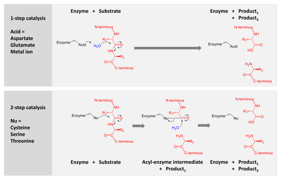
Catalytic Pathways
There are two primary mechanisms used to achieve catalysis:
- Direct Hydrolysis: Aspartic, glutamic, and metalloproteases activate a water molecule to perform a nucleophilic attack directly on the peptide bond.
- Covalent Intermediate: Serine, threonine, and cysteine proteases use a nucleophilic residue to covalently link the enzyme to the substrate. This creates an acyl-enzyme intermediate, which is subsequently hydrolyzed by water to release the final product and regenerate the enzyme.
Specificity and Regulation
Some proteases are promiscuous, meaning they can cleave a wide variety of proteins. Digestive enzymes like trypsin are examples; they typically recognize a single amino acid (such as Lysine or Arginine) rather than a long sequence. Others are highly specific, cleaving only a restricted set of sequences, which is common in viral proteases.
Regulation often occurs through autolysis, where a protease cleaves itself or other molecules of its own kind. This can either decrease activity (as seen in TEV protease) or activate the enzyme (as seen in the conversion of trypsinogen to trypsin).
Proteases Across Different Life Forms
| Organism/Entity | Primary Roles & Examples |
|---|---|
| Viruses | Cleaving massive polyproteins into functional units (e.g., Hepatitis C, Picornaviruses). |
| Bacteria | Nutrient recycling, protein quality control (AAA+ proteasome), and virulence factors (exotoxins). |
| Archaea | Cell signaling, metabolism, and quality control (LonB protease, 20S proteosome). |
| Plants | Developmental regulation and photosynthesis regulation. |
| Animals | Rapid signal amplification via cascades (blood clotting, complement system, apoptosis). |
Clinical Significance and Tumors
In humans, proteases play a dual role in cancer. Some facilitate tumor progression by degrading the extracellular matrix, allowing cancer cells to invade other tissues and metastasize. Conversely, more than 30 enzymes across three protease classes are known to have tumor-suppressive effects.
Protease Inhibitors
To prevent uncontrolled protein degradation, organisms use protease inhibitors. The serpin superfamily is a primary example, including antithrombin (which prevents excessive coagulation) and alpha 1-antitrypsin (which protects against inflammatory proteases).
In nature, some plants use inhibitors as defense mechanisms. For instance, raw soybeans contain trypsin inhibitors that discourage predators; these must be denatured through cooking to be safe for human consumption. In medicine, synthetic protease inhibitors are used as antiretroviral therapies to block the reproductive cycle of viruses like HIV/AIDS.
Frequently Asked Questions
What is the difference between an endopeptidase and an exopeptidase?
Endopeptidases cleave peptide bonds within the internal sequence of a protein, while exopeptidases only remove amino acids from the ends (terminals) of the protein chain.
How does a catalytic triad work?
A catalytic triad is a group of three amino acids—typically including a histidine—that work together to make another residue (like serine, cysteine, or threonine) highly nucleophilic, allowing it to attack the peptide bond of a substrate.
Why are protease inhibitors important in medicine?
They are used to treat viral infections, such as HIV/AIDS, by blocking the specific proteases the virus needs to replicate. They also exist naturally in the body to prevent enzymes from causing excessive inflammation or blood clotting.
What is the MEROPS database?
MEROPS is a comprehensive database used to classify proteases based on their evolutionary phylogeny, grouping them into clans and families based on structure and sequence similarity.
Can proteases be harmful?
Yes, some bacteria secrete proteases as exotoxins to destroy extracellular structures in a host, and certain proteases in tumors help cancer cells spread through the body by degrading the surrounding matrix.