Pelvis Anatomy: Structure, Function, and Sexual Dimorphism
The pelvis is a complex anatomical region located at the base of the trunk, serving as the critical transition zone between the abdomen and the thighs. It consists of both the pelvic region of the trunk and the embedded bony pelvis, which provides essential structural support and protects vital internal organs.
The pelvic region is composed of several distinct areas: the bony pelvis, the pelvic cavity (the space enclosed by the bones), the pelvic floor (the muscular diaphragm below the cavity), and the perineum, which is the area located below the pelvic floor.
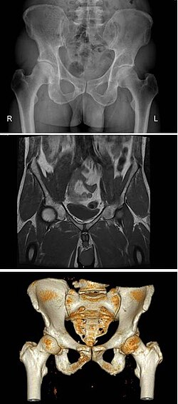
The Bony Pelvis: Structure and Composition
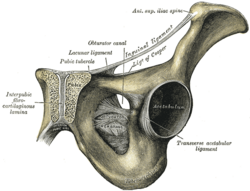
The pelvic skeleton, or bony pelvis, is divided into two primary components: the pelvic girdle and the pelvic spine. The pelvic girdle is formed by a pair of hip bones (os coxae), each consisting of three fused bones: the ilium, ischium, and pubis. These bones form a ring that connects the spine to the lower limbs.
The pelvic spine consists of the sacrum and the coccyx (tailbone). Together, these structures create a basin-shaped ring that anchors the vertebral column to the femora (thigh bones).
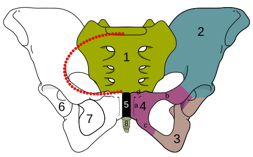
Anatomical Divisions of the Pelvis
The pelvis is further categorized by the pelvic brim, which divides the area into two main sections:
- Greater (False) Pelvis: The area located above the pelvic brim.
- Lesser (True) Pelvis: The area located below the pelvic brim, which contains the pelvic cavity.
The pelvic cavity is a body cavity bounded by the pelvic bones, primarily housing the rectum and reproductive organs. For clinical and obstetric purposes, this cavity is often analyzed across three planes: the inlet, the midplane, and the outlet.
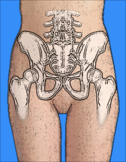
Mechanical Function and Muscular Support
Mechanically, the pelvis acts as a system of four twisted triangular rings. The superior rings are formed by the iliac bones, while the lower rings, formed by the rami of the pubic and ischial bones, support the acetabulum (the socket of the hip joint).
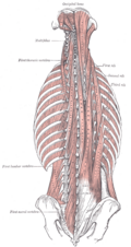
The pelvis also serves as a vital anchor for various muscle groups. The lumbosacral joint, connecting the sacrum to the last lumbar vertebra, is reinforced by the iliolumbar and lateral lumbosacral ligaments. This joint allows for flexion and extension, though axial rotation is not possible.
Key muscles associated with the pelvis include:
- Quadratus lumborum: Extends from the iliac crest to the 12th rib and lumbar vertebrae, assisting in lateral trunk bending and expiration.
- Iliopsoas: Comprising the psoas major and iliacus, this muscle group flexes and externally rotates the hip joints.
Sexual Dimorphism in the Pelvis
In mammals, the bony pelvis exhibits significant differences between males and females, primarily to accommodate childbirth. The gap in the middle of the pelvis is notably larger in females to allow offspring to pass through during birth.
Key Differences Between Male and Female Pelves
- Shape: The sides of the male pelvis converge from the inlet to the outlet, whereas the female pelvis remains wider.
- Subpubic Angle: In males, the angle between the inferior pubic rami is acute (approximately 70 degrees). In females, this is an obtuse angle (90–100 degrees), referred to as the pubic arch.
- Iliac Crests: Males typically have higher and more pronounced iliac crests, resulting in a deeper and narrower false pelvis.
The Caldwell–Moloy Classification
Developed in the 1930s by William Edgar Caldwell and Howard Carmen Moloy, this system classifies female pelves into four primary types based on the shape of the pelvic inlet:
| Pelvis Type | Inlet Shape | Characteristics | Obstetric Significance |
|---|---|---|---|
| Gynaecoid | Round or slightly oval | Wide subpubic arch, straight walls | Normal female pelvis; easiest for childbirth |
| Android | Wedge or heart-shaped | Prominent sacrum, triangular anterior segment | Reduced outlet; may cause birth difficulties |
| Anthropoid | Oval (greater anteroposterior diameter) | Straight walls, small subpubic arch | Deep and generally non-obstructed |
| Platypelloid | Flattened/Transversally wide | Short sacrum, reduced anteroposterior diameter | Associated with transverse arrest during birth |
Key Facts
- The pelvis connects the vertebral column to the femora.
- The pubic arch in females is obtuse (90–100°), while the subpubic angle in males is acute (70°).
- The gynaecoid pelvis is the most common type associated with uncomplicated childbirth.
- The pelvic girdle is composed of the ilium, ischium, and pubis.
- The pelvic floor acts as a muscular diaphragm supporting the pelvic cavity.
Frequently Asked Questions
What is the difference between the true and false pelvis?
The false (greater) pelvis is the area above the pelvic brim, while the true (lesser) pelvis is the area below the brim that contains the pelvic cavity and reproductive organs.
Which pelvic type is most common in women?
The gynaecoid pelvis is considered the normal female pelvis and was found in approximately 50% of specimens in the Caldwell-Moloy study.
What bones make up the pelvic girdle?
The pelvic girdle consists of two hip bones (os coxae), each formed by the fusion of the ilium, ischium, and pubis.
How does the male pelvis differ from the female pelvis?
The male pelvis is generally narrower, has a more acute subpubic angle, and possesses higher, more pronounced iliac crests compared to the wider, more open structure of the female pelvis.
What is the function of the pelvic floor?
The pelvic floor, or pelvic diaphragm, provides structural support for the pelvic cavity and the organs contained within it, separating the cavity from the perineum below.