Orbit Anatomy: Structure, Function, and Clinical Significance
In vertebrate anatomy, the orbit is the bony cavity or socket within the skull that houses the eye and its associated appendages. While the term often refers specifically to the bony structure, it can also encompass the entire contents of the socket. In a healthy adult human, the orbit has a total volume of approximately 28 milliliters, with the eyeball itself occupying about 6.5 milliliters of that space.
The orbital cavity is more than just a protective shell; it is a complex intersection of bone, muscle, nerves, and vasculature that ensures the eye can move precisely and transmit visual information to the brain.
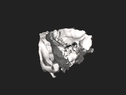
Key Facts
- Composition: Formed by a mosaic of seven distinct bones.
- Volume: Total orbital volume is ~28 ml; the eye occupies ~6.5 ml.
- Fragility: The medial wall (lamina papyracea) is the most delicate bone in the skull.
- Protection: The lateral wall is the thickest and most resistant to blunt force trauma.
- Critical Pathways: Contains the optic canal and superior orbital fissure for intracranial communication.
The Bony Structure of the Orbit
The orbits are shaped like four-sided pyramidal cavities that open toward the face and point backward into the skull. Each orbit consists of a base (the orbital margin), an apex, and four walls.
The Seven Bones of the Orbit
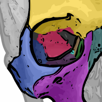
The orbital walls are not formed by a single bone but by a combination of seven embryologically distinct structures:
- Frontal bone: Forms the superior margin and the roof.
- Zygomatic bone: Forms the lateral wall and part of the inferior margin.
- Maxillary bone: Forms the floor and part of the medial wall.
- Sphenoid bone: Forms the posterior portion, including the optic canal (lesser wing) and the lateral posterior wall (greater wing).
- Ethmoid bone: Forms the medial wall; its extremely thin layer is known as the lamina papyracea.
- Lacrimal bone: Located in the medial wall and contains the nasolacrimal duct.
- Palatine bone: Contributes a minute portion to the orbital floor.
The Four Orbital Walls
- Roof (Superior Wall): Primarily the frontal bone and the lesser wing of the sphenoid. It contains the lacrimal fossa and trochlear fovea.
- Floor (Inferior Wall): Composed of the maxilla, zygomatic bone, and palatine bone. It features the infraorbital groove leading to the infraorbital foramen.
- Medial Wall: The thinnest wall, formed mainly by the ethmoid bone, maxilla, lacrimal bone, and sphenoid.
- Lateral Wall: The thickest and strongest wall, formed by the zygomatic bone and the greater wing of the sphenoid.
Openings and Communication Pathways
Several critical openings, known as foramina (windows) and fissures (grooves), allow nerves and blood vessels to enter and exit the orbit.
Major Foramina and Fissures
- Optic Canal: Transmits the optic nerve (Cranial Nerve II) and the ophthalmic artery.
- Superior Orbital Fissure: A major communication pathway containing cranial nerves III, IV, and VI (which control eye movement) and the ophthalmic branches of the trigeminal nerve (V1).
- Supraorbital Foramen: Contains the supraorbital nerve, a branch of the frontal nerve.
- Infraorbital Foramen: Contains the infraorbital nerve, a branch of the maxillary nerve (V2).
- Inferior Orbital Fissure: Connects the orbit to the pterygopalatine and infratemporal fossae, carrying the infraorbital artery and vein.
These openings are clinically significant because they can serve as pathways for infections or cancer to spread from the orbit into the brain or deep facial structures.
Orbital Contents and Function
The primary function of the orbit is to hold and protect the eye. However, it also houses the complex machinery required for vision and movement.
Muscles and Vasculature
Eye movement is managed by six extraocular muscles: the superior, inferior, medial, and lateral rectus muscles, and the superior and inferior oblique muscles. Blood supply is provided by the ophthalmic artery, which is particularly important as it can provide collateral blood flow to the brain during internal carotid infarcts via the circle of Willis. Deoxygenated blood is drained by the superior ophthalmic vein.
The Lacrimal System
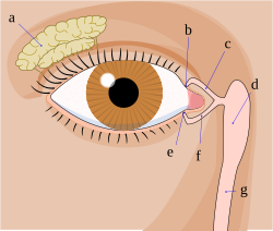
The orbit contains the lacrimal gland, which produces tears. These tears drain through the lacrimal sac and the nasolacrimal duct into the nasal cavity.
| Component | Primary Structures | Key Function/Feature |
|---|---|---|
| Bony Walls | 7 bones (Frontal, Zygomatic, Maxilla, Sphenoid, Ethmoid, Lacrimal, Palatine) | Structural protection of the globe |
| Nerves | Cranial Nerves II, III, IV, V, VI | Vision and ocular motility |
| Muscles | 4 Rectus, 2 Oblique muscles | Precise eye movement |
| Drainage | Lacrimal gland, sac, and nasolacrimal duct | Tear production and drainage |
| Vessels | Ophthalmic artery and superior ophthalmic vein | Blood supply and drainage |
Clinical Significance
Because the orbit is a confined space, any increase in volume can lead to exophthalmos (protrusion of the eye).
- Graves' Ophthalmopathy: Associated with Graves' disease, this involves the buildup of extracellular matrix proteins and fibrosis in the rectus muscles, causing the eye to bulge forward.
- Tumors: Gliomas or meningiomas of the optic nerve can cause axial protrusion. Enlargement of the lacrimal gland typically pushes the eye inferiorly and medially.
- Trauma: Due to its extreme thinness, the lamina papyracea of the medial wall is one of the most common sites of fracture during orbital trauma.
Frequently Asked Questions
What is the most fragile part of the eye socket?
The most fragile part is the lamina papyracea, a very thin section of the ethmoid bone that forms part of the medial wall. It is frequently fractured during orbital trauma.
Which nerves are responsible for moving the eye?
Eye movement is controlled by cranial nerves III (oculomotor), IV (trochlear), and VI (abducens), which pass through the superior orbital fissure to reach the extraocular muscles.
What causes the eye to protrude forward (exophthalmos)?
Exophthalmos occurs when excessive tissue accumulates behind the eye. Common causes include Graves' disease (due to muscle fibrosis), tumors such as gliomas or meningiomas, or inflammation of the lacrimal gland.
Why is the ophthalmic artery clinically important beyond the eye?
The ophthalmic artery can act as a critical collateral pathway to the circle of Willis, providing an alternative blood source to the brain in cases where the internal carotid artery is blocked.
How many bones make up the human orbit?
The human orbit is composed of seven bones: the frontal, zygomatic, maxillary, sphenoid, ethmoid, lacrimal, and palatine bones.