Miosis: Causes, Symptoms, and the Physiology of Pupil Constriction
Miosis, also known as myosis or "pinpoint pupil," is the medical term for the excessive constriction of the pupil. Derived from the Ancient Greek word múein, meaning "to close the eyes," miosis occurs when the opening of the iris narrows, limiting the amount of light that enters the eye. To understand miosis, it is helpful to distinguish it from two related conditions: mydriasis, which is the dilation (widening) of the pupil, and anisocoria, a condition where the two pupils are of unequal size.
Key Facts
- Miosis is the excessive constriction of the pupil.
- It can be caused by natural factors (bright light), aging, specific diseases, or drug exposure.
- The process is controlled by the parasympathetic nervous system via the oculomotor nerve.
- Opioids are a well-known pharmacological cause of pinpoint pupils.
- The opposite of miosis is mydriasis.
The Physiology of the Photomotor Reflex
The resizing of the pupil is governed by the photomotor reflex, a complex biological process that regulates light intake. When light enters the eye, it strikes three types of photoreceptors in the retina: the rods and cones (responsible for image formation) and photosensitive ganglion cells. While rods and cones react quickly, the ganglion cells react more sluggishly and provide information regarding ambient light levels.
These ganglion cells perform several critical functions, including the regulation of pupil size, the entrainment of circadian rhythms, and the suppression of the hormone melatonin. The process follows a specific neural pathway: retinal photoreceptors convert light into electric impulses, which travel to the pretectal nucleus of the high midbrain, bypassing the primary visual cortex and lateral geniculate nucleus.
From the pretectal nucleus, neurons send axons to the Edinger-Westphal nucleus. Visceromotor axons then travel along both the left and right oculomotor nerves (Cranial Nerve III). These axons synapse on ciliary ganglion neurons, whose parasympathetic axons ultimately innervate the iris sphincter muscle, causing it to contract and produce miosis.
Common Causes of Miosis
Miosis can result from a variety of triggers, ranging from normal physiological responses to severe medical emergencies.
Natural and Age-Related Factors
The most common cause of miosis is exposure to bright light pointed directly at the eye. Additionally, senile miosis refers to the natural reduction in pupil size that occurs as a person ages.
Medical Conditions and Diseases
Several pathological states can lead to constricted pupils, including:
- Neurological and Vascular Issues: Hemorrhage into the pons (intracranial hemorrhage), Horner's syndrome, and fatal familial insomnia.
- Pain and Inflammation: Cluster headaches (often accompanied by ptosis, or drooping eyelids), hemicrania continua, and iridocyclitis.
- Genetic and Other Disorders: Ehlers Danlos Syndrome, hereditary disorders, and aphakia (the absence of the lens of the eye).
Pharmacological and Chemical Triggers
Many drugs and chemicals can induce miosis by affecting the nervous system or the muscles of the iris:
- Opioids: Fentanyl, morphine, heroin, and methadone (though pethidine is a notable exception).
- Cholinergic Agents: Acetylcholine, acetylcholinesterase inhibitors, and parasympathomimetics like pilocarpine eye drops.
- Psychotropic Medications: Antipsychotics (such as risperidone, haloperidol, chlorpromazine, olanzapine, and quetiapine), mirtazapine (a NaSSA antidepressant), and certain MAO inhibitors.
- Other Substances: Nicotine products (cigarettes, gum, chewing tobacco), imidazolines (clonidine, naphazoline, oxymetazoline, tetrahydrozoline), serotonin antagonists (such as ondansetron/Zofran), and certain cancer chemotherapy drugs (camptothecin derivatives).
- Toxins: Organophosphates and, in rare instances, exposure to mustard gas.
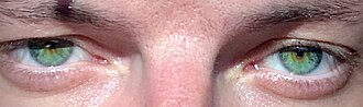
Summary of Miosis Characteristics
| Term | Definition | Primary Mechanism/Cause |
|---|---|---|
| Miosis | Excessive pupil constriction | Parasympathetic activation / Iris sphincter contraction |
| Mydriasis | Pupil dilation | Sympathetic activation / Iris dilator muscle |
| Anisocoria | Unequal pupil size | Neurological dysfunction or localized injury |
| Senile Miosis | Age-related constriction | Natural aging process |
Frequently Asked Questions
What is the difference between miosis and mydriasis?
Miosis is the constriction or narrowing of the pupil, while mydriasis is the dilation or widening of the pupil.
Which opioids are known to cause pinpoint pupils?
Opioids such as morphine, heroin, fentanyl, and methadone typically cause miosis; however, pethidine is a notable exception to this effect.
How does light cause the pupil to constrict?
Light triggers photosensitive ganglion cells in the retina, which send signals through the pretectal nucleus to the Edinger-Westphal nucleus. This activates the oculomotor nerve and the iris sphincter muscle, resulting in miosis.
Can medications other than opioids cause miosis?
Yes. Miosis can be caused by antipsychotics, cholinergic agents, certain antidepressants like mirtazapine, nicotine products, and some chemotherapy drugs.
What is anisocoria?
Anisocoria is the medical condition where one pupil is more dilated than the other, rather than both being constricted or dilated equally.